XB-IMG-134968
Xenbase Image ID: 134968
|
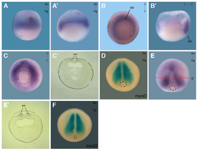
Fig. 2. zcchc24 expression from blastula to gastrula stages. Whole mount in situ hybridization using DIG labelled zcchc24 (A-Câ, E,Eâ) or fluorescein labelled myoD (D,F) was performed on embryos from blastula to gastrula stages. (A,Aâ) At blastula stages, zcchc24 is expressed as a ring around the marginal zone in both dorsal and ventral sides. The embryos display the animal hemisphere to the top. (B,Bâ) In the beginning of gastrulation (st 10.5), zcchc24 expression is restricted to the dorsal mesoderm in both involuting and non-involuting marginal zone. (C,Câ, E,Eâ) At late gastrula (st 11 and 12), zcchc24 transcripts are detected in two lateral mesoderm stripes around the blastopore that culminate in the dorsal mesoderm, the somitogenic mesoderm, but is excluded from the midline. (D,F) myoD is expressed in the somitogenic mesoderm. (Aâ , Bâ) Hemisections of (A,B), respectively. (Câ, Eâ) Transverse sections of st 11 and st 12 embryos, respectively, with dorsal side displayed to the top. (B,C) Vegetal views. (D,E,F) Dorsal views. Dashed lines delimitate the blastopore. An, animal; Vg, vegetal; D, dorsal; V, ventral; dbl, dorsal blastopore lip; sm, somitogenic mesoderm. Image published in: Vitorino M et al. (2014) Copyright © 2014. Image reproduced with permission of the Publisher.
Image source: Published Larger Image Printer Friendly View |