Click here to close Hello! We notice that you are using Internet Explorer, which is not supported by Xenbase and may cause the site to display incorrectly. We suggest using a current version of Chrome, FireFox, or Safari.
XB-IMG-135547

Xenbase Image ID: 135547


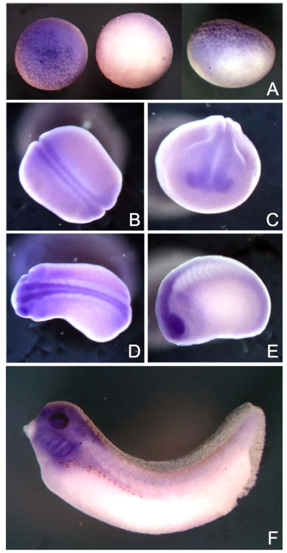
Fig. 2. Spatial expression patterns of xMBD3 transcripts in Xenopus embryos on whole-mount in situ hybridization. (A) Animal pole (left), vegetal pole (middle), and lateral (right) views at stage 8. Dorsal (B) and anterior (C) views of neural groove embryos at stage 19. Dorsal (D) and lateral (E) views of tailbud embryos at stage 23. (F) Lateral view of a tadpole stage embryo.

Image published in: Iwano H et al. (2004)

Copyright © 2004. Image reproduced with permission of the Publisher, Elsevier B. V.

GeneSynonymsSpeciesStage(s)Tissue
mbd3.SLOC108712329, MGC69548, xmbd3X. laevisThroughout NF stage 19optic vesicle
neural plate
pre-chordal neural plate
chordal neural plate
mbd3.SLOC108712329, MGC69548, xmbd3X. laevisThroughout NF stage 23eye
optic vesicle
cranial neural crest
head
neural tube
posterior neural tube
anterior neural tube
mbd3.SLOC108712329, MGC69548, xmbd3X. laevisThroughout NF stage 33 and 34eye
pharyngeal arch
mandibular arch
hyoid arch
branchial arch
brain
forebrain
midbrain
hindbrain
otic vesicle
mbd3.SLOC108712329, MGC69548, xmbd3X. laevisThroughout NF stage 8animal hemisphere
animal

Image source: Published

Larger Image
Printer Friendly View

Return to previous page