XB-IMG-135858
Xenbase Image ID: 135858
|
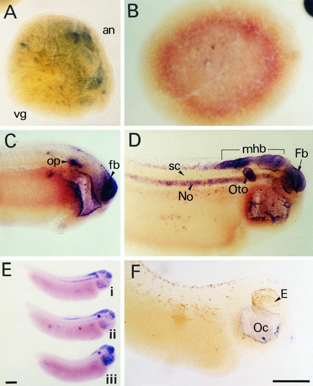
FIG. 5. Spatial distribution of Xrel3
mRNA during development. Albino
embryos were subjected to whole mount
in situ hybridization analysis using Xrel3-
specific probes. The dark blue staining
indicates localization of message. A, blastula;
B, gastrula; C, late neurula; D and
E, larva; F, control; op, otic placode; fb,
forebrain; mhb, mid-hindbrain; oto, otocyst;
No, notochord; sc, spinal cord; E, eye;
Oc, oral cavity. Scale bar 5 0.5 mm. Image published in: Yang S et al. (1998) Copyright © 1998. Image reproduced with permission of the Publisher.
Image source: Published Larger Image Printer Friendly View |