XB-IMG-1359
Xenbase Image ID: 1359
|
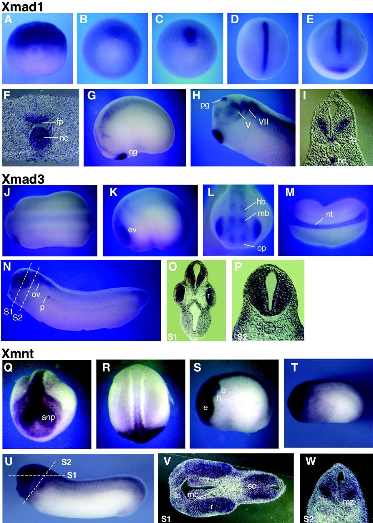
Figure 3. Spatial expression pattern of Xmad1, Xmad3, and Xmnt in Xenopus embryos. (A-I) Xmad1, (J-P) Xmad3, and (Q-W) Xmnt. (A) Stage 6.5 lat., (B) stage 10 veg., (C) stage 11 veg., (D) stage 13 d., (E) stage 17 ant., (F) stage 17, transversal section, (G) stage 19, (H) stage 27, (I) stage 27, transversal section, (J) stage 20 d., (K) stage 20 lat., (L) stage 23 ant., (M) stage 23 d., (N) stage 29, (O) transversal section S1 of N, (P) transversal section S2 of N, (Q) stage 19 ant., (R) stage 19 d., (S) stage 20 lat., (T) stage 23 lat., (U) stage 27, (V) stage 27 sagittal section S1, and (W) stage 27 transversal section S2. anp, anterior neural plate; b, branchial crest segment; cg, cement gland; d, dorsal; ev, eye vesicle; fb, forebrain; fp, floor plate; h, hyoid crest segment; hb, hindbrain; hc, hypochord; lat, lateral; mz marginal zone; nc, notochord; nt, neural tube; veg, vegetal; mb, midbrain; op, olfactory placode; otic vesicle; P, pronephros; pg, pineal gland; sc, spinal chord; r, retina; V, trigeminal ganglion; VII, geniculate ganglion. The dashed line indicates the plane of the section. Image published in: Juergens K et al. (2005) Copyright © 2005. Image reproduced with permission of the Publisher, John Wiley & Sons.
Image source: Published Larger Image Printer Friendly View |