XB-IMG-139809
Xenbase Image ID: 139809
|
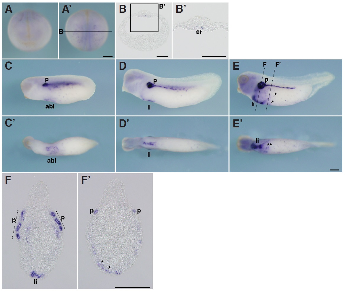
Fig.2. In situ hybridization analysis of igfbp1 during X. tropicalis development. (A,Aâ) Stage 20, (A) Anterior view (Aâ) dorsal view. (B,Bâ) Transversal section of an embryo at stage 20. (Bâ) Magnified view of the boxed area in (B). Dorsal side is displayed towards the top. Igfbp1 is expressed in part of the archenteron roof near the dorsal midline. (C,Câ) Stage 25, (D,Dâ) stage 30, (E,Eâ) stage 35, (C,D,E) lateral view, (Câ,Dâ,Eâ) ventral view. (F,Fâ) Transversal section of an embryo at stage 35. Positions of sections are indicated by black lines, and letters mark corresponding panels. ar, archenteron roof; abi, anterior blood islands; li, liver; p, pronephric tubule. Arrowheads indicate scattered blood-like cells. Scale bars indicate 200 mm. Image published in: Haramoto Y et al. (2014) Copyright © 2014. Image reproduced with permission of the Publisher, University of the Basque Country Press.
Image source: Published Larger Image Printer Friendly View |