XB-IMG-139812
Xenbase Image ID: 139812
|
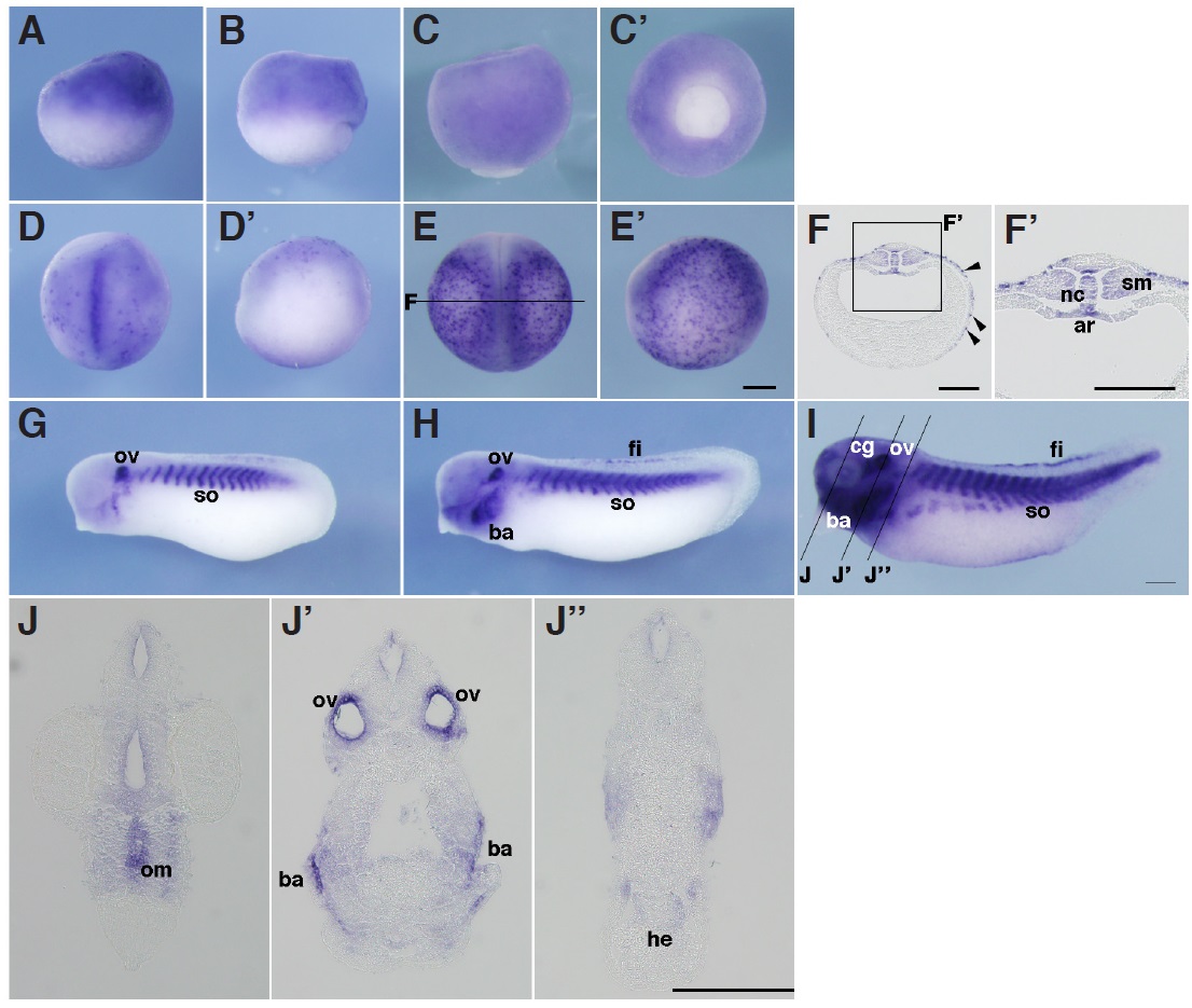
Fig. 5. In situ hybridization analysis of igfbp5 during X. tropicalis development. (A) Stage 9, (B) stage 10.5, (A,B) lateral view, (C,Câ) stage 12, (C) lateral view, (Câ) vegetal view, (D,Dâ) stage 15, (E,Eâ) stage 20, (D,E) dorsal view, (Dâ,Eâ) lateral view, (F,Fâ) transversal section of an embryo at stage 20. (Fâ) Magnified view of the boxed area in (F). (G) Stage 25, (H) stage 30, (I) stage 35, (J,Jâ,Jââ) transversal section of an embryo at stage 35. Positions of sections are indicated by black lines, and letters mark corresponding panels. nc, notochord; ar, archenteron roof; sm, somatic mesoderm; ov, otic vesicle; so, somite; ba, branchial arch; fi, fin; cg, cranial ganglia; om, oral membrane; ov, otic vesicle; he, heart. Scale bars indicate 200 mm. Image published in: Haramoto Y et al. (2014) Copyright © 2014. Image reproduced with permission of the Publisher, University of the Basque Country Press. Larger Image Printer Friendly View |