Click here to close
Hello! We notice that you are using Internet Explorer, which is not supported by Xenbase and may cause the site to display incorrectly.
We suggest using a current version of Chrome,
FireFox, or Safari.
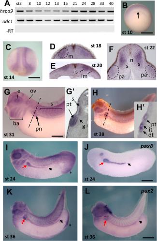
Figure 1. Expression pattern of Hspa9 in Xenopus laevis embryo. A: RT-PCR hspa9 expression in Xenopus laevis embryos from stages 3 to 40 of development with odc1 as house-keeping gene. BâH: Whole-mount ISH of hspa9. B: Early gastrula, arrow indicates the position of the blastoporal lip. C: Early neurula stage (anterior view, dorsal side up) shows hspa9 expression in neural and mesodermal territories. D,E: hspa9 is expressed in somite (s), mesoderm (m) and intermediate mesoderm (im). F: hspa9 labels the pronephros anlage (pa). G: At early tail bud stage, strong expression of hspa9 in eye (e), otic vesicle (ov), branchial arches (ba), somites and pronephros, from proximal (red arrow) to distal part (black arrow). Gâ²: Section through the plan of dotted line in G. H,Hâ²: Tadpole stage shows persistent hspa9 expression in the pronephros tubule. IâL: hspa9 pronephros expression was compared with pax8 and pax2 renal markers. Arrows show similar hspa9 and pax expression domains, up to the tip of the nephric duct. A strong hspa9 expression is present in the proctodeum (asterisk in I, K). Abbreviations: dt, distal tubule; g, glomus; it, intermediate tubule; n, notochord; pt, proximal tubule. Scale bars = 0.5 mm in BâH; 1 mm in IâL.Download figure to PowerPoint