XB-IMG-151886
Xenbase Image ID: 151886
|
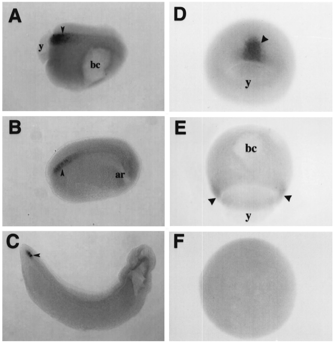
Fig. 5. Spatial expression of integrin a2 and a3 mRNAs. Whole-mount in situ hybridization was performed on albino Xenopus embryos using antisense
digoxygenin-labeled a2 (AâC) or a3 (DâF) transcripts. (A) Stage 11.5 gastrula, anterior to the right, dorsal to the top with staining of a2 in posterior
notochord (arrowhead). (B) Stage 19 neurula with a2 staining localized to the posterior end of the notochord (arrowhead). (C) Slightly oblique dorsal view of
a stage 32 tailbud embryo. a2 expression persists in the posterior tip of the notochord (arrowhead). (D) Dorsal lip view of a normal stage 10.5 control
gastrula. The cells expressing a3 are the dorsal involuting mesoderm (arrowhead). (E) Side view of a gastrula stage embryo that has been dorsalized by
exposure to LiCl. Arrowheads indicate the circumblastoporal expression of a3 encircling the yolk plug. (F) Side view of an embryo that has been ventralized
by UV irradiation of the vegetal hemisphere. Embryos treated with UV light lack detectable a3 staining. Labeled sense transcripts for both a2 and a3 were
included as specificity controls in parallel hybridizations for each experiment (data not shown). y, yolk plug; bc, blastocoel; ar, archenteron. Image published in: Meng F et al. (1997) Copyright © 1997. Image reproduced with permission of the Publisher, Elsevier B. V.
Image source: Published Larger Image Printer Friendly View |