XB-IMG-151912
Xenbase Image ID: 151912
|
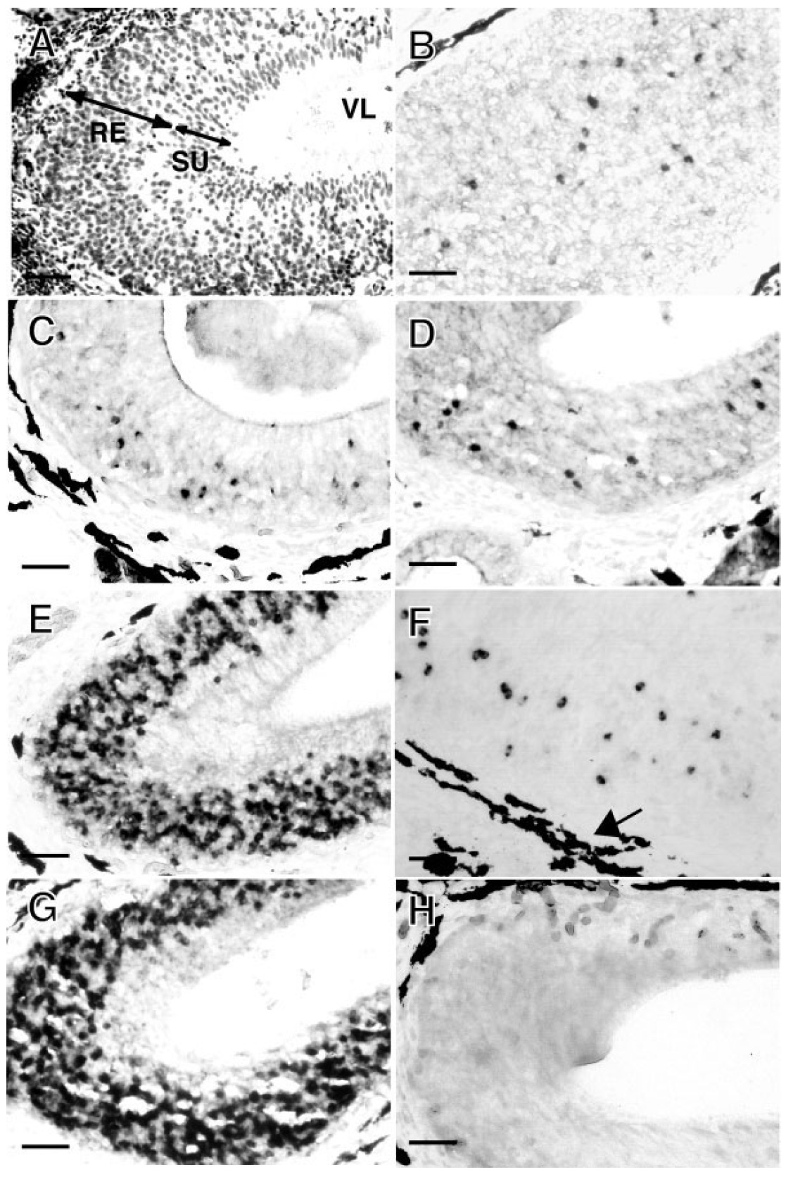
Fig. 3. Analysis of Xenopus V2R expression in the vomeronasal
organs by in situ hybridization. Cross sections of Xenopus vomeronasal
epithelium were stained with hematoxylin (A). The cross
sections were hybridized to digoxigenin-labeled antisense
probes which were derived from clones A-1 (B), B-1 (C), C-1 (D),
E-1 (E), xV2R1 (F), a mixture of A-1, C-1, and E-1 (G), and sense
probes derived from a mixture of A-1, C-1, and E-1 (H). RE, receptor
cell layer; SU, supporting cell layer; VL, lumen of the VNO. Black
spots around the outside of the VNO in AâH (for example, arrow
in F) are melanocyte aggregates. Scale bars 50 m. Image published in: Hagino-Yamagishi K et al. (2004) Copyright © 2004. Image reproduced with permission of the Publisher.
Image source: Published Larger Image Printer Friendly View |