XB-IMG-152459
Xenbase Image ID: 152459
|
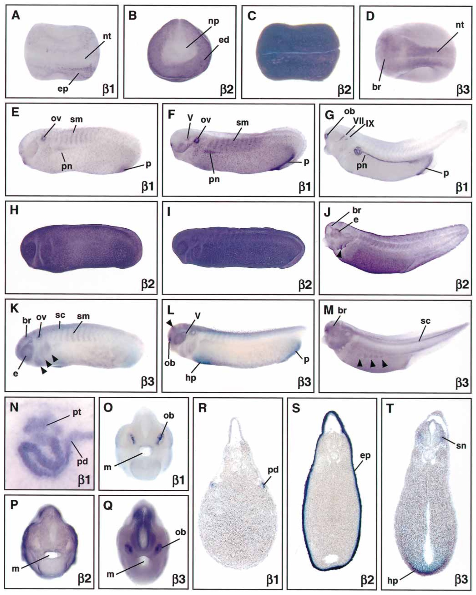
Fig. 2 Expression of Na,K-ATPase b subunit genes during Xenopus
embryogenesis. Whole-mount in situ hybridizations were per-
formed on Xenopus embryos with antisense probes for b1, b2, and
b3 subunits. Sections of stage 29 (S), stage 30 (T) and stage 37 (R)
embryos stained in whole-mount were cut at 30â40 mm. Dorsal
(A, C, D) and lateral (E-N) views are shown with anterior to the
left. Frontal views (B, O-Q) and sections (R-T) are oriented with
dorsal to the top. A Stage 19 embryo with punctate b1 expression
in the epidermis. B, C b2 expression was confined to the non-neural
ectoderm at stage 15 (B) and throughout the epidermis at stage 19
(C). D Stage 19 embryo showing b3 expression in the developing
brain and neural tube. E-G b1 transcripts appeared at stage 26
(E) initially in the otic vesicles, pronephric anlage, somites, and
proctodeum. Later, at stage 29 (F) staining was noticeable in the
trigeminal (V) nerves. By stage 37 (G), b1 transcripts were also
evident in the olfactory bulbs, facial (VII) and glossopharyngeal
(IX) nerves. H-J Extensive epidermal expression of b2 transcripts
was observed in stage 25 (H), stage 29 (I), and stage 38 (J) embryos.
Arrowhead indicates high levels of b2 in the gills. Note the increasing
b2 expression in the brain and eyes. K-M b3 gene expression
was found at stage 26 (K) in the brain, eyes, otic vesicles, somites,
and cranial neural crest (arrowheads). By stage 30 (L), strong stain
ing was evident at the forebrain-midbrain junction (arrowhead), in
the region of the hepatic primordium, the proctodeum, and olfac-
tory bulbs. b3 transcripts were seen at stage 38 laterally in the
migrating abdominal muscle anlagen (arrowheads). N High magni
fication of a stage 37 embryo illustrating b1 expression in the
pronephric kidney. O-Q Frontal views showing b1 (O), b2 (P), and
b3 (Q) expression in the developing head. R-T Transverse sections
through the midtrunk of stage 37 (R) and at the level of the pro
nephros of stage 29 (S) and stage 30 (T) embryos. b1 transcripts
(R) were present in the pronephric duct, b2 expression was confined
to the epidermis, and b3 was observed in spinal nerves and the
hepatic primordium. Abbreviations: br, brain; e, eye; ed, ectoderm;
ep, epidermis; hp, hepatic primordium; m, mouth, np, neural plate;
nt, neural tube; ob, olfactory bulb; ov, otic vesicle; p, proctodeum;
pd, pronephric duct; pt, pronephric tubules; pn, pronephros; sc,
spinal cord; sm, somites; sn, spinal neurons.
Image published in: Eid SR and Brändli AW (2001) Copyright © 2001. Image reproduced with permission of the Publisher and the copyright holder. This is an Open Access article distributed under the terms of the Creative Commons Attribution License.
Image source: Published Larger Image Printer Friendly View |