XB-IMG-153795
Xenbase Image ID: 153795
|
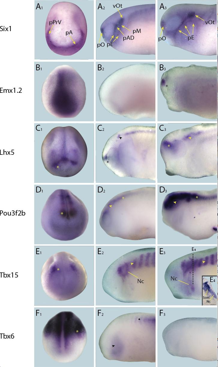
Figure 4âfigure supplement 1.
Expression of targets not expressed in placodes in whole-mount Xenopus embryos.
Expression dynamics for each target are shown across a range of developmental stages: A1âF1 show expression in neural plate stage embryos, A2âF2 show early tail bud stage embryos and A3âF3 show late tail bud stage embryos. (A) Expression of PPE marker gene Six1 is shown as reference for placodal domains (for details see Pandur and Moody, 2000; Schlosser and Ahrens, 2004). (B) Emx1.2 is expressed broadly in the neural plate in neural plate stages (B1), and becomes restricted to the forebrain in late tail bud stages (B3; asterisk). (C) Lhx5 is expressed in the forebrain at all developmental stages (C1âC3; asterisk), and at early and late tail bud stages Lhx5 is also expressed in the hindbrain and spinal cord (C3; arrowhead). (D) Pou3f2b is expressed in the neural plate and developing neural tube (D1; asterisk) at neural plate stages. Expression in the brain and spinal cord is maintained during early and late tail bud stages (D2 and D3 ; arrowhead and asterisk, respectively). (E) Tbx15 is expressed in a restricted domain of the anterolateral neural folds in neural plate stages (E1; asterisk). At tail bud stages expression is prominent in somites (E2 and E3; arrowhead) and migrating neural crest cells of the hyoid and first branchial neural crest streams (Nc). Both of these expression domains are maintained into late tail bud stages (E3 and E4). E4 shows section at the level indicated in E3 (dotted line). Bar in E4: 100 μm. (F) Throughout all developmental stages (F1âF3) Tbx6 is expressed strongly in the posterior paraxial and lateral plate mesoderm (F1 and F2; asterisk) with weaker expression in the pharyngeal arches (F2; arrowhead). Subsequently, itâs expressed in somites, as indicated by a diamond in F2. Abbreviations: pA: anterior placodal region; pAD: anterior lateral line placode; pE: epibranchial placode; pM: middle lateral line placode; pO: olfactory placode; vOt: otic vesicle; pPrV: profundal/trigeminal placodes. Image published in: Riddiford N and Schlosser G (2016) © 2016, Riddiford et al. Creative Commons Attribution license
Image source: Published Larger Image Printer Friendly View |