XB-IMG-157988
Xenbase Image ID: 157988

|
Fig. 4.
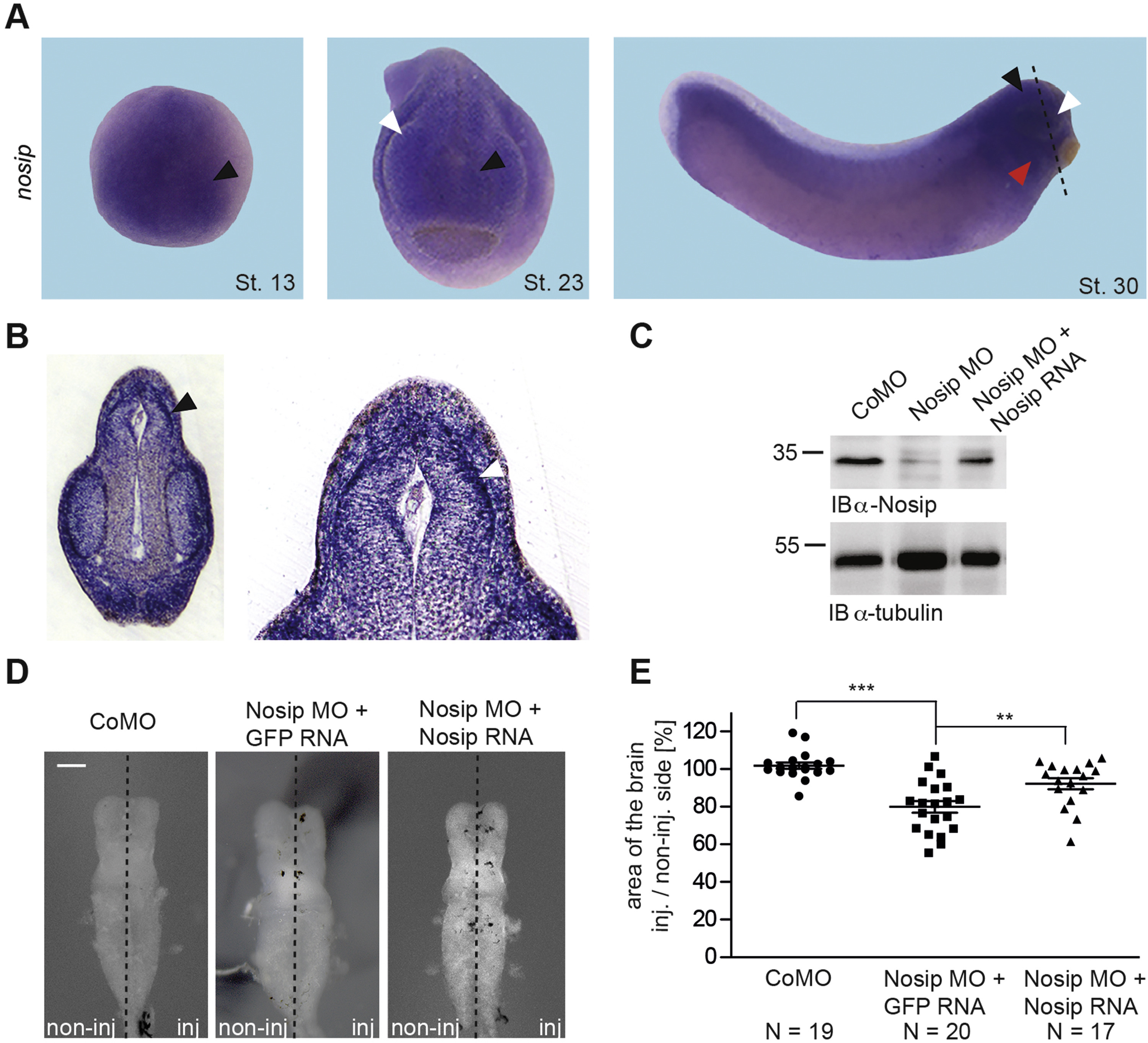
Nosipis expressed in the developing neural tissue ofXenopusand knockdown of Nosip causes microcephaly (A) Expression analysis of Nosip by WMISH at indicated Xenopus laevis developmental stages; white arrowheads point to eyes, black arrowheads to anterior neural tissue (St.13) or brain (St. 23/St. 30), and red arrowhead to pharyngeal arches (B) Transversal vibratome section of the embryo head with arrowheads pointing to differentiated neurons of the brain; the dotted line in A indicates the level of the section shown in B. (C) Immunoblot analysis of Xenopus embryo lysates using a Nosip-specific antiserum; immunoblot with an α-tubulin-specific antibody served as loading control; lysates were generated from Xenopus embryos at stage 20 after bilateral injection of 23 ng Nosip MO or Control MO together with 0.5 ng human NOSIP mRNA in 2-cell stage Xenopus embryos as indicated (D) Bright field images of Xenopus brains at stage 42, anterior to the top after unilateral injection of Nosip MO or Control MO and human NOSIP mRNA as indicated; scale bar 200 µm (E) Statistical evaluation of the data shown in D, N = number of independently evaluated brains. Image published in: Hoffmeister M et al. (2017) Copyright © 2017. Image reproduced with permission of the Publisher, Elsevier B. V.
Image source: Published Larger Image Printer Friendly View |
