XB-IMG-171053
Xenbase Image ID: 171053
|
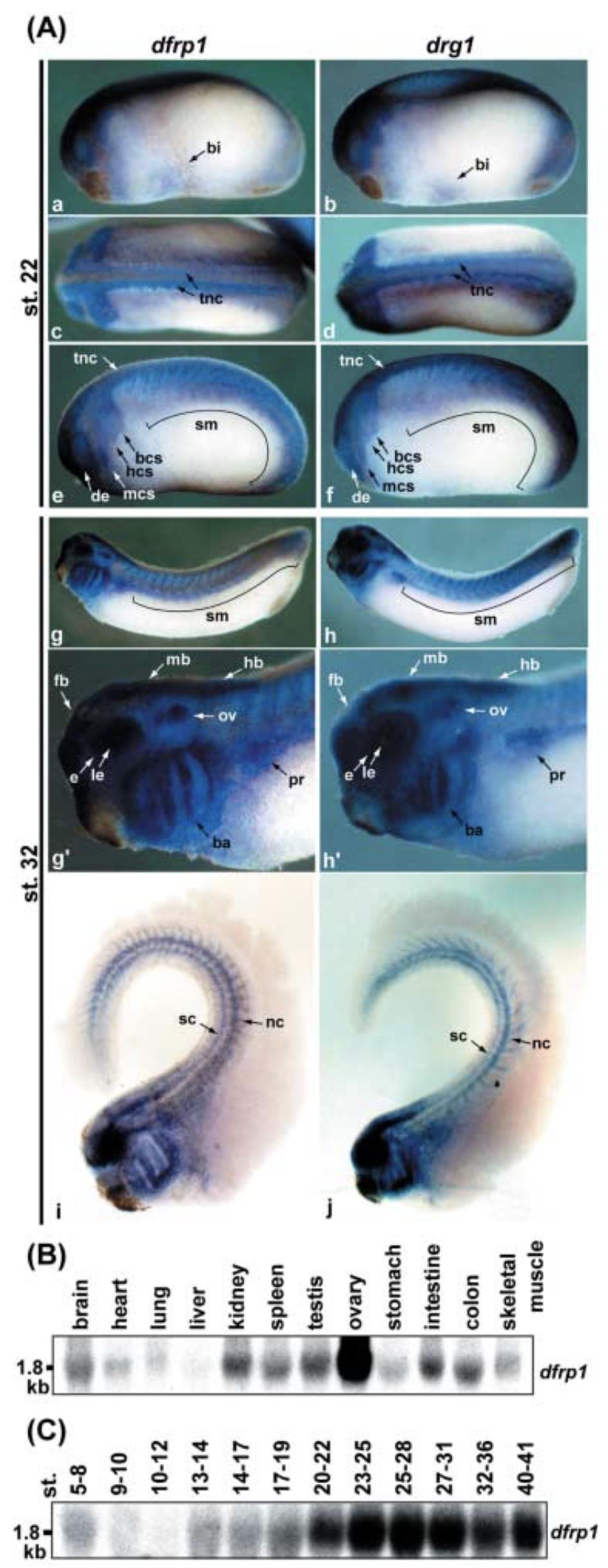
Figure 6 Expression analysis of dfrp1 and drg1 in X. laevis. (A) Spatial expression of dfrp1 and drg1 transcripts during Xenopus embryonic development. (a,b) Ventral views, anterior left; (c,d) Dorsal views, anterior left; (eâj) Lateral views, anterior left; (gâ² and hâ²) Higher magnification images of the anterior part of the embryo depicted in g and h, respectively; (i, j) Clearing of embryo by benzyl alcohol:benzyl benzoate (2 : 1). Abbreviations: ba, branchial arch; bcs, branchial crest segment; bi, blood islands; de, developing eyes; e, eyes; fb, forebrain; hb, hindbrain; hcs, hyoid crest segment; le, lens; mb, midbrain; mcs, mandibular crest segment; nc, notochord; ov, otic vesicle; pr, pronephros; sc, spinal cord; sm, somite; tnc, trunk neural crest. (B) Tissue-specific expression of dfrp1 mRNAs in adult Xenopus.Total RNAs were isolated from the indicated adult tissues for Northern blotting. (C) Temporal expression of dfrp1 transcripts during X. laevis embryonic development.Total RNAs were isolated from Xenopus embryos at the indicated stages for Northern blotting. In (B,C), the same membrane that was used in our previous paper (Ishikawa et al. 2003) was reprobed.Therefore, quality and amount of RNA loaded were confirmed. Image published in: Ishikawa K et al. (2005) Copyright © 2005. Image reproduced with permission of the Publisher.
Image source: Published Larger Image Printer Friendly View |