XB-IMG-173621
Xenbase Image ID: 173621
|
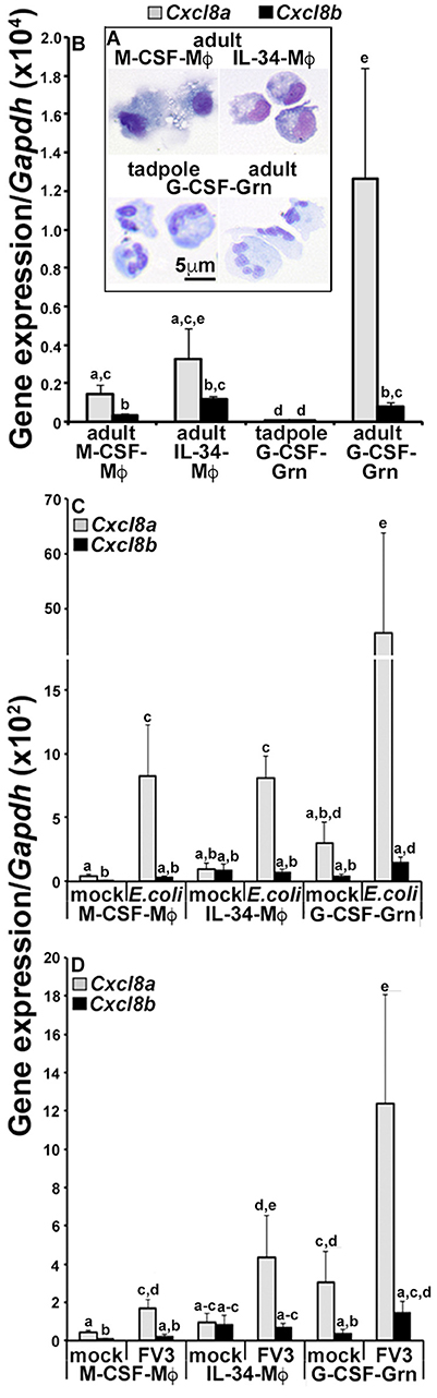
Figure 3. Analyses of CXCL8a and CXCL8b gene expression in frog macrophages and granulocytes. (A) Cytology of tadpole rG-CSF-elicited peritoneal granulocytes (G-CSF-Grn) and adult frog bone marrow-derived macrophages, differentiated with rM-CSF (M-CSF-MÏ) or rIL-34-MÏs (L-34-MÏ) and granulocytes (Grn), differentiated with rG-CSF (G-CSF-Grn). (B) The adult frog M-CSF-MÏ, IL-34-MÏ, and G-CSF-Grn were examined for their steady state gene expression of Cxcl8a and Cxcl8b. (C) The adult frog M-CSF-MÏ, IL-34-MÏ, and G-CSF-Grn were either mock infected (saline) or challenged with FV3 at a multiplicity of infection of 0.5 PFU/cell. After 24 h of challenge, cells were examined for Cxcl8a and Cxcl8b gene expression relative to Gapdh (N = 5). (D) M-CSF-MÏ, IL-34-MÏ, and G-CSF-Gran were mock challenged (saline) or challenged with heat-killed E. coli for 24 h before Cxcl8a and Cxcl8b gene expression analysis, relative to Gapdh (N = 5). All results are presented as means + SEM. Above-head letters denote statistical designations: experimental groups described by distinct letters are statistically different (P < 0.05) while those marked by the same letters are not. Image published in: Koubourli DV et al. (2018) Copyright © 2018 Koubourli, Yaparla, Popovic and Grayfer. Creative Commons Attribution license
Image source: Published Larger Image Printer Friendly View |