XB-IMG-175265
Xenbase Image ID: 175265
|
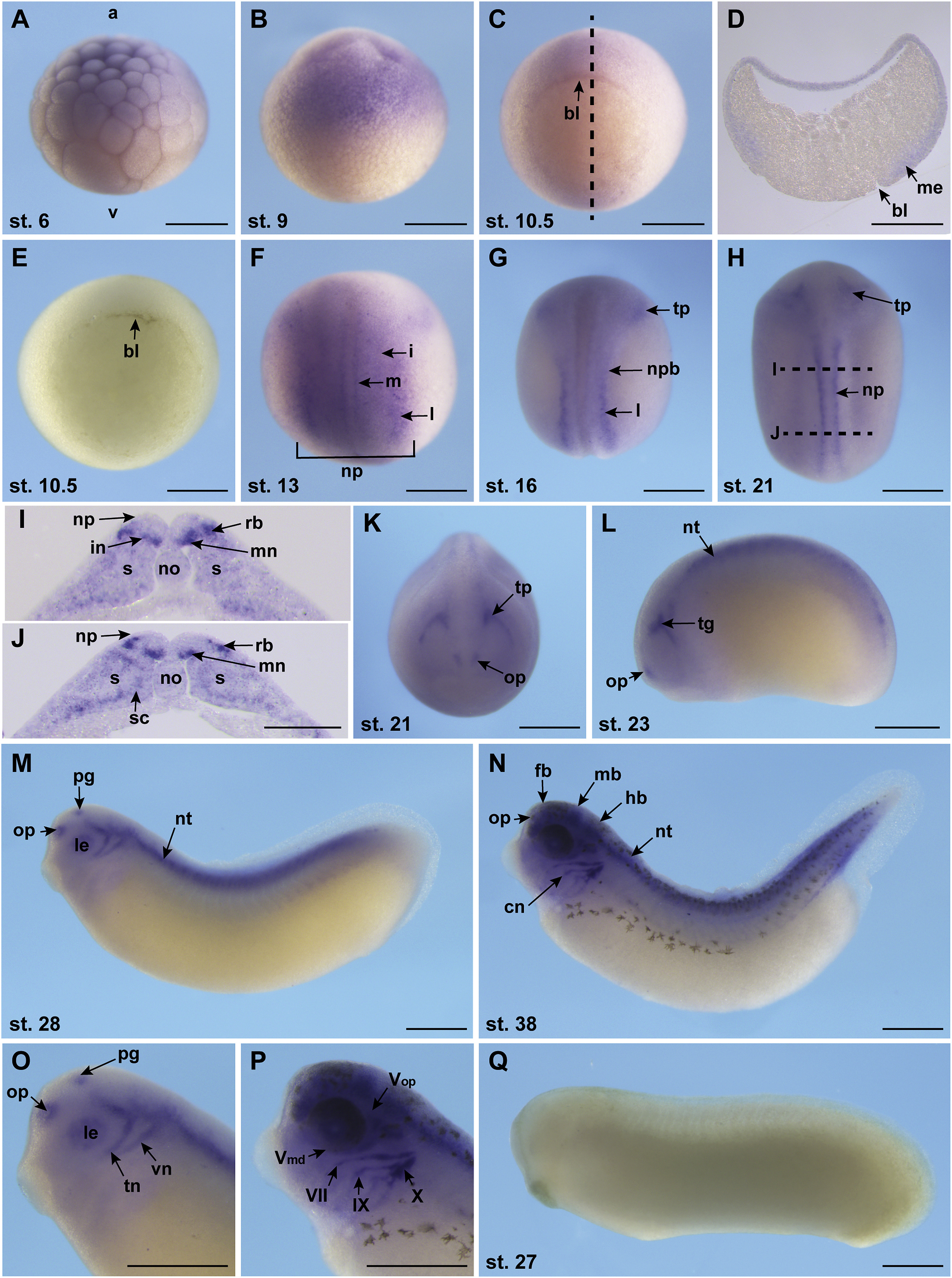
Fig. 5. Spatial gene expression pattern of kalrn. Kalrn gene expression was analyzed by whole mount in situ hybridization. Embryonic stages are indicated. The detailed expression pattern is described in the text. A Embryo at cleavage stage, lateral view. B Blastula stage embryo, lateral view. C Gastrula stage embryo, blastopore lip is indicated. D Transverse section of embryo in C; section plane is indicated in C. E Sense control of gastrula stage embryo. F-H,K Neurula stage embryos, dorsal view. I,J Transverse sections of embryo shown in H. Section planes are indicated in H. K Anterior view of embryo shown in H. M-N Tailbud stage embryos, lateral view. O,P Higher magnification of the embryo heads shown in M,N, respectively; the mandibular branch (Vmd) and the ophthalmic branch (Vop) of the trigeminal nerve (V), the facial VII, the glossopharyngeal (IX), and the vagus (X) nerve are indicated in P. Q Control in situ hybridization using a sense trio probe; tailbud stage embryo. Abbreviations: a: animal; bl: blastopore lip; cn: cranial nerves; fb: forebrain; hb: hindbrain; i: intermediate domain of primary neurons; in: interneuron; l: lateral domain of primary neurons; le: lens; m: medial domain of primary neurons; mb: midbrain; me: mesoderm; mn: motor neuron; no: notochord; np: neural plate; npb: neural plate border; nt: neural tube; op: olfactory placode; pg: pineal gland; rb: Rohon-Beard neuron; s: somites; sc: sclerotome; tg: trigeminal ganglion; tn: trigeminal nerve; tp: trigeminal placode; v: vegetal; vn: vestibulo cochlear nerve. Scale bar: 500â¯Î¼m (I,J: 200â¯Î¼m). Image published in: Kratzer MC et al. (2019) Copyright © 2019. Image reproduced with permission of the Publisher, Elsevier B. V.
Image source: Published Larger Image Printer Friendly View |