XB-IMG-180381
Xenbase Image ID: 180381
|
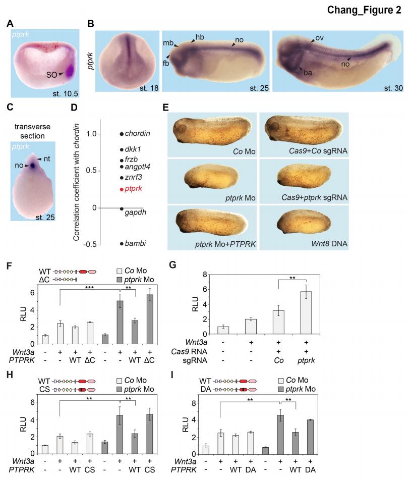
Figure 2 | Ptprk inhibits Wnt signaling in the Spemann organizer (A-C) In situ hybridization of ptprk in Xenopus tropicalis at (A) gastrula (hemisected, dorsal to the right), (B) neurula, tailbud, and tadpole stages, and in (C) transverse dissected tailbud embryo. ba, branchial arches; fb, forebrain; hb, hindbrain; mb, midbrain; no, notochord; nt, neural tube; ov, otic vesicle; SO, Spemann organizer. (D) Data mining using data from Ding et al. (2017), showing gene expression correlation with a dorsal/organizer marker chordin. Xenopus dkk1, frzb, and angptl4 are known organizer-expressed genes, gapdh is shown as housekeeping gene, and bambi is a ventrally expressed gene. (E) Representative phenotypes of tailbud stage Xenopus tropicalis embryos injected animally at 2- to 8- cell stage and as indicated. For quantification, see Figure 2 - figure supplement 1D-E. (F-I) Topflash reporter assays performed with neurulae (stage 18). Embryos were injected animally at 2- to 8-cell stage (F, H-I) or one cell stage (G) with reporter plasmids and the indicated mRNAs and Mos. Domain structures of WT PTPRK and mutants are shown on top. Normalized Topflash activity of Co930 injected embryos only with reporter plasmids was set to 1. Data in all graphs are displayed as means ± SD, and show one representative of multiple independent experiments with three biological replicates. RLU, relative light units. ** P < 0.01, ***P < 0.001. Image published in: Chang LS et al. (2020) © 2020, Chang et al. Creative Commons Attribution license
Image source: Published
Larger Image Printer Friendly View |