XB-IMG-180384
Xenbase Image ID: 180384
|
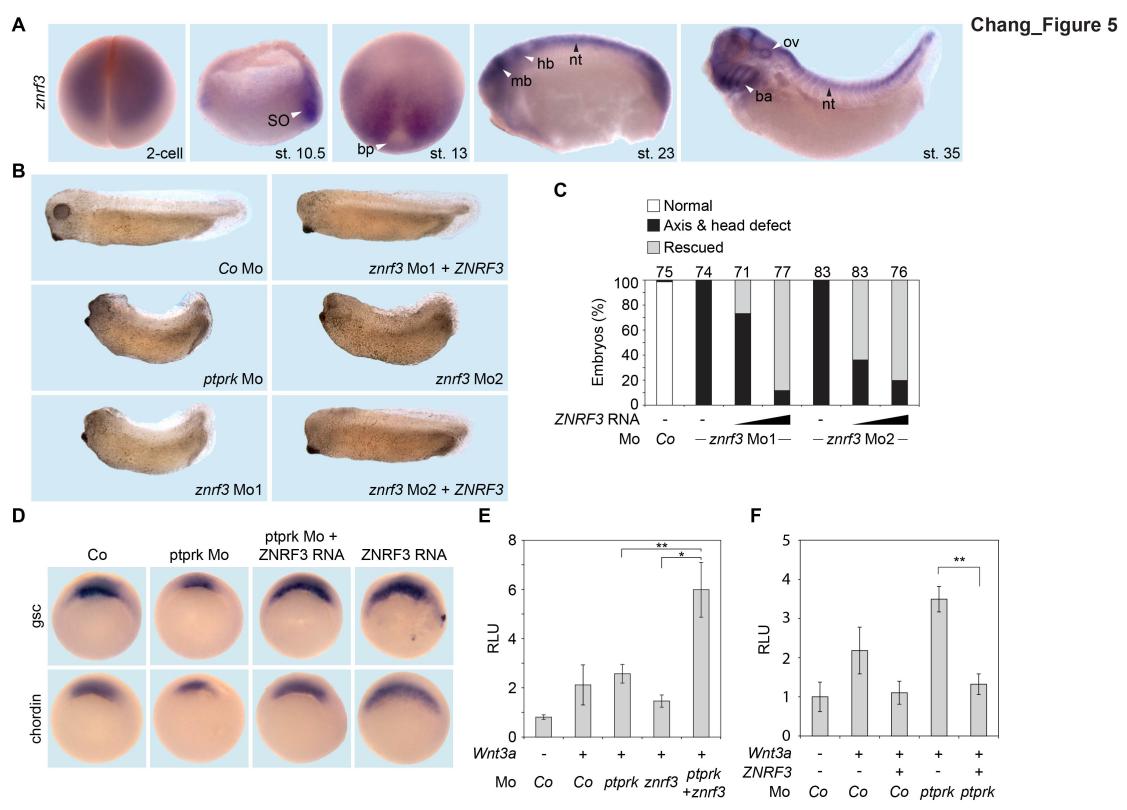
Figure 5 | Znrf3 is coexpressed- and cooperates with Ptprk in early Xenopus embryos (A) Spatial expression of znrf3 in Xenopus tropicalis embryos at blastula (animal view), gastrula (hemisected dorsal to the right), neurula, tailbud and tadpole stages. ba, branchial arches; bp, blastopore; hb, hindbrain; mb, midbrain; nt, neural tube; ov, ovic vesicle; SO Spemann organizer. (B) Representative phenotypes of tailbud stage Xenopus tropicalis embryos injected animally at 2- to 8- cell stage as indicated. (C) Quantification of phenotypes shown in (B). The number of embryos per condition is indicated on the top. (D) Whole mount in situ hybridization of gsc and chordin in gastrula embryos (stage 10.5). Embryos were injected at 2- to 8-cell stage animally with Co or ptprk Mo with or without ZNRF3 RNA. For quantification, see Figure 5 â figure supplement 1C. (E) Topflash reporter assay performed with neurulae (stage 18). Embryos were injected animally at 2- to 8-cell stage as indicated. Suboptimal dosages of ptprk or znrf3 Mos were used in this experiment. Normalized Topflash activity of Co Mo injected embryos was set to 1. (F) Topflash reporter assay performed with neurulae (stage 18). Embryos were injected animally at 2- to 8-cell stage as indicated. Normalized Topflash activity of Co Mo injected embryos was set to 1. Data in all graphs are displayed as means ± SD, and show one representative of multiple independent experiments with three biological replicates. RLU, relative light units. ** P < 0.01. Image published in: Chang LS et al. (2020) © 2020, Chang et al. Creative Commons Attribution license
Image source: Published Larger Image Printer Friendly View |