XB-IMG-190301
Xenbase Image ID: 190301
|
||||||||||
|
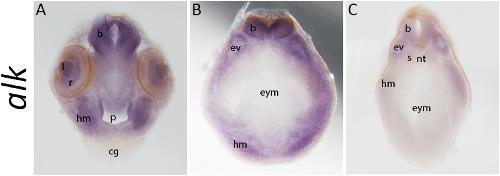
Fig. 3. alk is expressed in head structures of Xenopus laevis tadpoles. Coronal sections of Xenopus laevis tadpoles at stage 37, after in situ hybridisation, reveal internal structures with alk expression (Nieuwkoop and Faber, 1994). Anterior to posterior (AâC) 150 μm sections at the levels of the optic cup (A), ear vesicle (B) and somites (C) (r = retina, l = lens, p = pharynx, b = brain, hm = head mesenchyme, cg = cement gland, nt = notochord, s = somite, ev = ear vesicle, eym = endodermal yolk mass). Image published in: Moreno MM et al. (2021) Copyright © 2021. Creative Commons Attribution license
Image source: Published Larger Image Printer Friendly View |