XB-IMG-191234
Xenbase Image ID: 191234
|
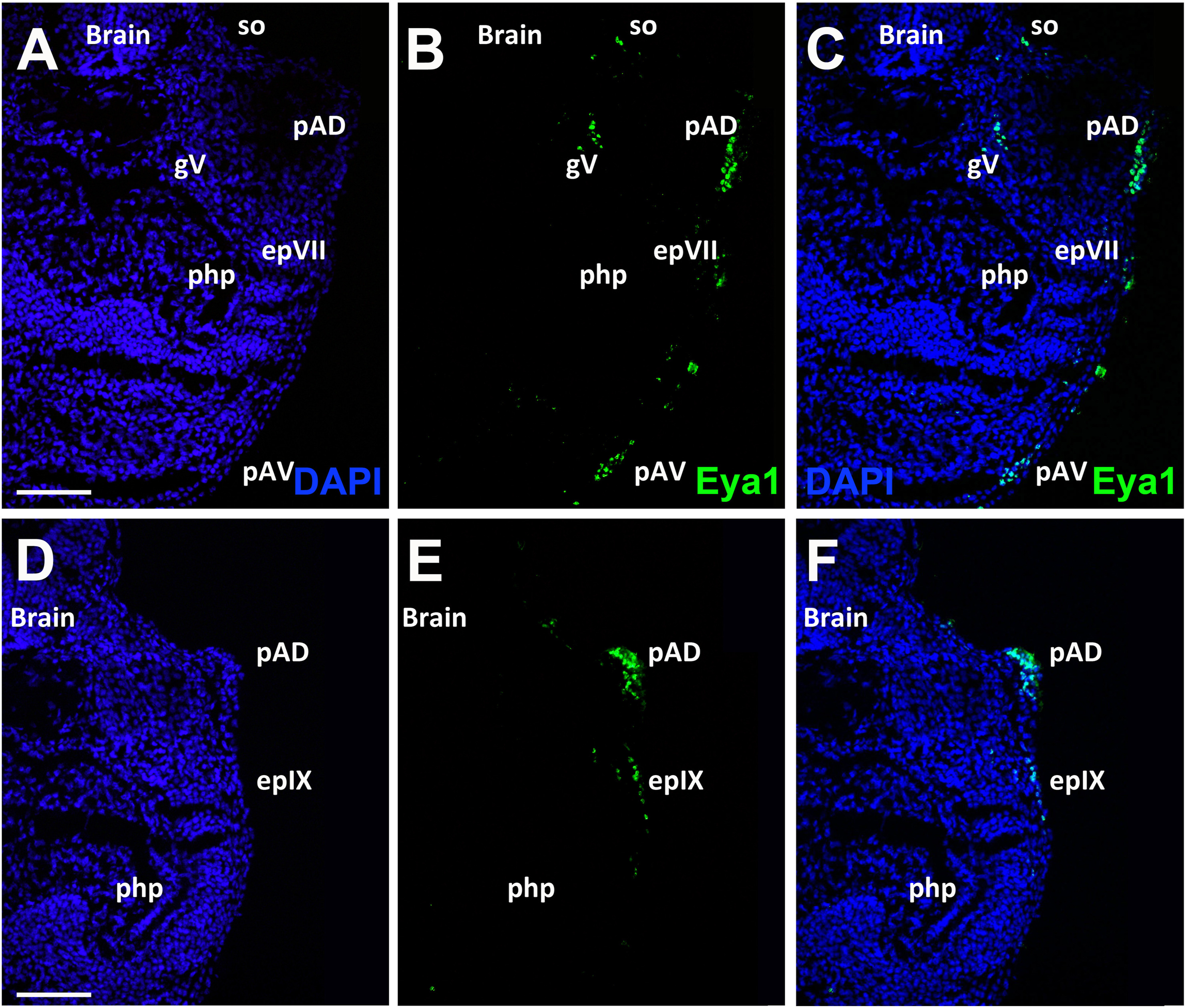
Fig. 12. Eya1 distribution in transverse sections of Xenopus laevis at stage 40 (cont.). A-F: Overview of stage 40 tadpole showing nuclear DAPI staining (first column), Eya1 immunostaining (second column) and merged channels (third column). Eya1 is expressed in trigeminal ganglion (gV), anterodorsal lateral line placode (pAD), anteroventral lateral line placode (pAV), sensory ridge of supraorbital lateral line (so), the facial epibranchial placode (epVII) and glossopharyngeal epibranchial placode (epIX). Eya1 expression is undetectable in brain and pharyngeal pouches (php). Scale bars: A, D: 100 μm (for A-C and D-F). Image published in: Almasoudi SH and Schlosser G (2021) Copyright © 2021. Image reproduced with permission of the Publisher, Elsevier B. V. Larger Image Printer Friendly View |