XB-IMG-192699
Xenbase Image ID: 192699
|
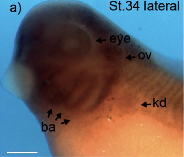
Fig.6.Ba GID genes are expressed in ciliated organs during X. laevis development. Spatial analysis of expression of GID complex subunits rmnd5a (A) and mkln1 (B) in X. laevis. WMISH of WT X. laevis embryos at developmental stage 34 (St. 34; lateral views in Aa and Ba) and the corresponding transverse (b,e,d) and sagittal (c) sections (ov, otic vesicle; pe, prosencephalon; ba, branchial arches; kd, pronephric kidney; gc, ganglion cells). Images are representative of three experiments. Images in Ab and Bb are the result of tiling multiple fields of view. Scale bars: 300 μm in Aaâc and Baâc; 100 μm in Ad, Ae, Bd and Be. This image is extracted from figure published in: Hantel F et al. (2022), Image published in: Hantel F et al. (2022) Copyright © 2022. Image reproduced with permission of the Publisher, The Company of Biologists Ltd.
Image source: Published Larger Image Printer Friendly View |