XB-IMG-200206
Xenbase Image ID: 200206
|
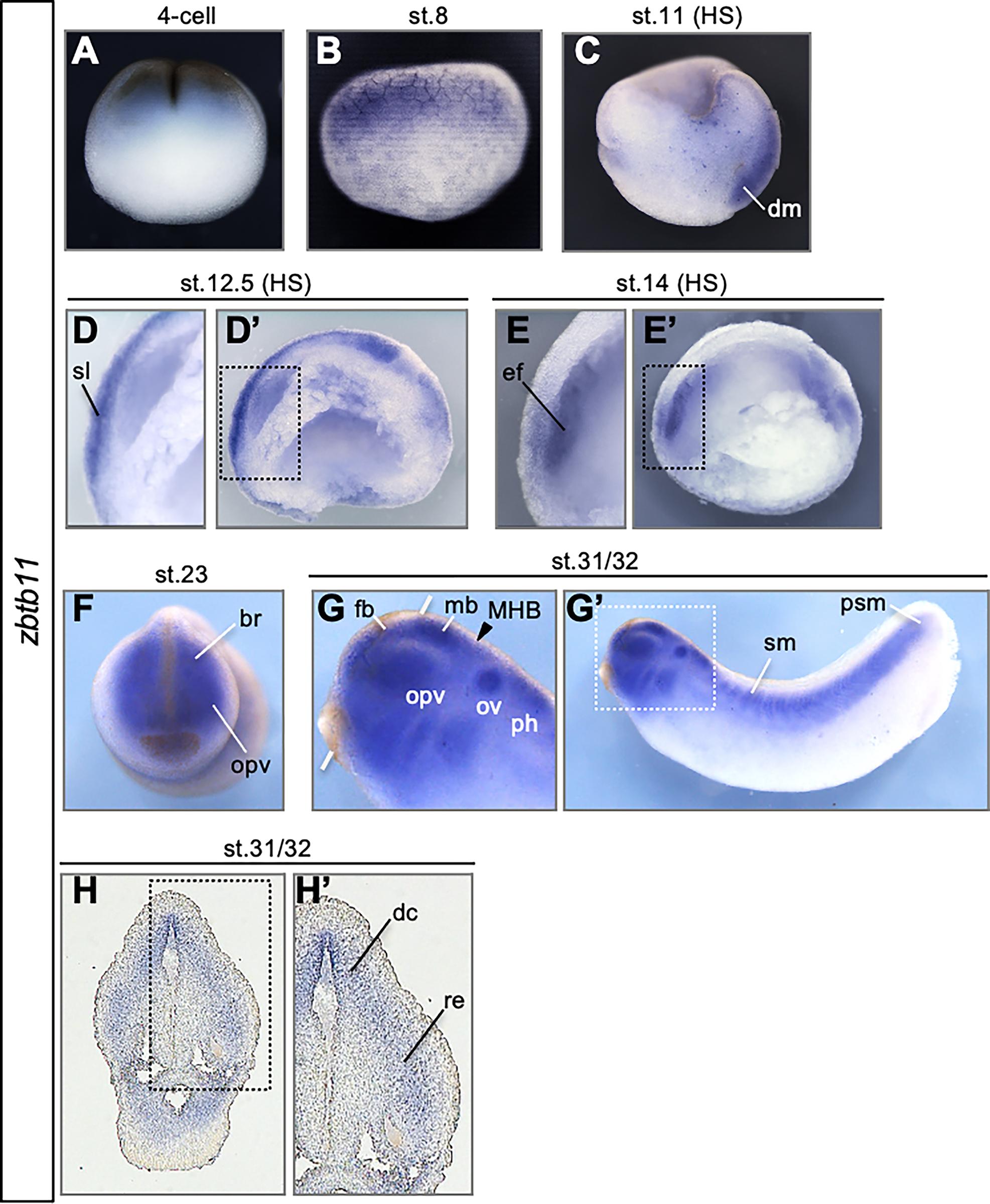
Fig 1. Developmental expression patterns of zinc finger and BTB domain-containing 11 (zbtb11).
(A-G) Expression of zbtb11 analyzed by whole-mount in situ hybridization (WISH) using whole embryos (A,B,F,G) or sagittal hemisections (C,D,E; indicated by HS). (H) Transverse cryosection following WISH. Developmental stages (st.) are indicated at the top of each panel. Lateral view with animal pole side up (A-C). Lateral view with dorsal side up (D,E,G). Anterior view with dorsal side up (F,H). Dashed boxes (D’,E’,G’,H’) indicate enlarged images in D, E, G, and H. An arrowhead indicates the position of the midbrain and hindbrain boundary (MHB), and white thick lines indicate the level of transverse sections in H (G). br, brain; dc、diencephalon, dm, dorsal mesoderm; ef, eye field; fb, forebrain; mb, midbrain; MHB, midbrain and hindbrain boundary; opv, optic vesicles; ov, otic vesicles; ph, posterior part of the hindbrain; psm, presomitic mesoderm; re, retina; sl, sensorial layer; sm, somites.
https://doi.org/10.1371/journal.pone.0293852.g001 Image published in: Satou-Kobayashi Y et al. (2024) © 2024 Satou-Kobayashi et al. Creative Commons Attribution license
Image source: Published Larger Image Printer Friendly View |