XB-IMG-2104
Xenbase Image ID: 2104
|
|||||||||||||||||||||||||||||||||||
|
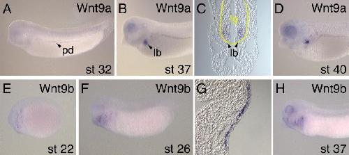
Figure 4. In situ hybridization analysis of Wnt9a and Wnt9b expression during Xenopus embryonic development. A: Lateral view of stage 32 embryo showing Wnt9a transcripts in the pronephric duct (pd). B: Lateral view of stage 37 embryo showing expression of Wnt9a in the pronephric duct and lung bud (lb) C: Transverse section through the anterior foregut of a stage 37 embryo showing staining of Wnt9a in the endodermal cells of the lung buds. D: Lateral view of the head region of a stage 40 embryo showing expression of Wnt9a in the eye, lung bud, and pronephric duct. E: Lateral view of stage 22 embryo showing expression of Wnt9b in the branchial arch region. F: Lateral view of stage 26 embryo showing persistent expression of Wnt9b in the branchial arch region. G: Transverse section through the branchial arch region of a stage 26 embryo showing Wnt9b expression confined to the inner ectodermal layer of the epidermis. H: Lateral view of a stage 37 embryo showing expression of Wnt9b in the branchial arches, the eye, and adjacent to the cement gland. GenBank accession numbers for Xenopus tropicalis Wnt9a and Wnt9b are DQ658159 and DQ658160, respectively. Image published in: Garriock RJ et al. (2007) Copyright © 2007. Image reproduced with permission of the Publisher, John Wiley & Sons.
Image source: Published Larger Image Printer Friendly View |