XB-IMG-25029
Xenbase Image ID: 25029
|
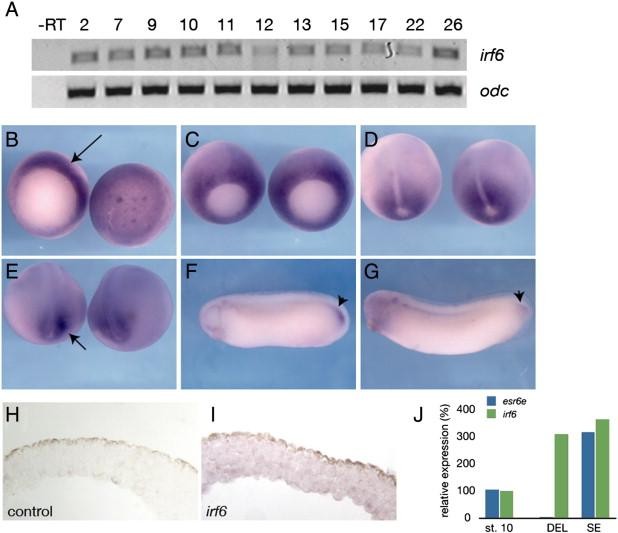
Fig. 5. Expression of irf6 in Xenopus laevis. (A) RT-PCR analysis of irf6 expression at different stages of development. Developmental stages are indicated at the top (Nieuwkoop and Faber, 1956). â RT, negative control reaction without reverse transcriptase. (BâG) Whole mount in situ hybridization for irf6. (B) Stage 10.5, left is vegetal view, arrow indicates expression in the dorsal marginal zone. Right is animal pole view. (C) Stage 11, expression is throughout marginal zone, but beginning to be lost from dorsal midline. (D) Stage 12.5, expression around blastopore, absent from prospective notochord. (E) Stage 18, expression flanking prospective tailbud region, arrow. (F) Stage 28 and (G) stage 30, notable expression in tailbud, arrowheads. (H) Section of animal cap hybridized with an irrelevant control probe (xnr3). (I) Section of animal cap hybridized with an antisense irf6 probe. Diffuse expression is found in both superficial and deep layers. (J) Quantitative RT-PCR of irf6 (green) and esr6e (blue) expression in ectodermal explants; st. 10 (stage 10 whole embryo), DEL (deep layer explant), SE (superficial layer explant). Image published in: Sabel JL et al. (2009) Copyright © 2009. Image reproduced with permission of the Publisher, Elsevier B. V.
Image source: Published Larger Image Printer Friendly View |