XB-IMG-26142
Xenbase Image ID: 26142
|
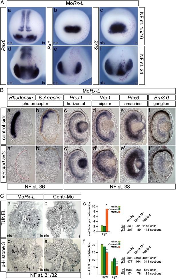
Fig. 3. Rx-L function is needed after eye field induction. (A, B) WMISH analysis of staged embryos injected with MoRx-L at 4-cell stage into one of the dorsoanimal blastomeres. (AaâAf) Frontal views, injected side to the right. At NF stage 15/16, the expressions of Pax6 (Aa), Rx1 (Ab) and Six3 (Ac) are not significantly affected due to suppression of Rx-L function. (AdâAf) At tailbud stage (NF stage 24), the expressions of Pax6 (Ad), Rx1 (Ae) and Six3 (Af) are all reduced in the injected sides. (Baâf') Transversal gelatin/albumin sections of injected embryos probed with the markers of different retinal cell types, with dorsal side upward. The staining of each probe in the retina of injected side (Ba'âf') should be compared with that in the corresponding control side of the same embryo (Baâf) respectively. The red dashes in Ba' mark the presumptive RPE. Suppression of Rx-L function led to a dramatically reduced expression of the photoreceptor markers, Rhodopsin (Ba, Ba') and Arrestin-C (Bb, Bb'), and a broadened expression of the bipolar cell maker, Vsx1 (Bd, Bd') and the amacrine/ganglion cell marker, Pax6 (Be, Be'). The expression levels Prox1 (Bc, Bc') and Brn3.0 (Bf, Bf') remain almost unchanged upon MoRx-L injection. (C) Effects of MoRx-L microinjection on apoptosis and cell proliferation. (Ca, b, d, e) Transversal sections (dorsal sides up and injected sides to the right) of embryos injected with 2.5pmol of MoRx-L (Ca, d) or Contr-Mo (Cb, e) subjected to TUNEL assay (Ca, b) or immunostaining of phosphorylated histone H3 (p-Histone 3) (Cd, e) at NF stage 31/32 to detect apoptotic or proliferating cells, respectively. (Cc, f) Graphs showing the numbers of TUNEL positive cells (Cc) or p-Histone 3 positive cells (Cf) in total or eye area of the non-injected side (green bars), Contr-Mo injected side (green bars), and MoRx-L (orange bars). The average of TUNEL or p-Histone-3 positive cell numbers on per section was determined in each embryo. In the TUNEL assay, for non-injected, n = 5 embryos; for Contr-Mo, n = 2 embryos; for MoRx-L, n = 3 embyros. In the p-Histone 3 detection, for non-injected, n = 5 embryos; for Contr-Mo, n = 2 embryos; for MoRx-L, n = 3 embyros. Values are given as means ± S.E.M. p = 0.28, , p < 0.05, compared with the non-injected side (Student's t-test). Quantification of the counted cells and sections is shown next to the graphs respectively. Image published in: Wu HY et al. (2009) Copyright © 2009. Image reproduced with permission of the Publisher, Elsevier B. V.
Image source: Published Larger Image Printer Friendly View |