Click here to close Hello! We notice that you are using Internet Explorer, which is not supported by Xenbase and may cause the site to display incorrectly. We suggest using a current version of Chrome, FireFox, or Safari.
XB-IMG-40632

Xenbase Image ID: 40632
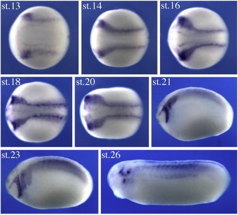

Fig. 1. Myo10 is expressed in neural crest tissues during Xenopus development. Myo10 expression is first observed in Xenopus embryos at late gastrula stages, at the borders of the forming neural plate, where the neural crest will arise. Expression subsequently increases in the cranial neural crest and continues as they migrate towards the branchial arches. By tailbud stages, the expression decreases, mainly remaining in the cranial ganglia. All embryos are oriented with anterior to the left.

Image published in: Nie S et al. (2009)

Copyright © 2009. Image reproduced with permission of the Publisher, Elsevier B. V.

GeneSynonymsSpeciesStage(s)Tissue
myo10.2.SLOC108702915, myosin-X, MyosinXX. laevisThroughout NF stage 13 to NF stage 20neural crest
neural plate border
cranial neural crest
neuroectoderm
myo10.2.SLOC108702915, myosin-X, MyosinXX. laevisThroughout NF stage 21 to NF stage 23paraxial mesoderm
cranial neural crest
mandibular crest
hyoid crest
branchial crest
migratory neural crest cell
trunk neural crest
myo10.2.SLOC108702915, myosin-X, MyosinXX. laevisThroughout NF stage 26myotome
dorsal fin
trunk somite
cranial ganglion
cranial placode
somite

Image source: Published

Larger Image
Printer Friendly View

Return to previous page