Click here to close Hello! We notice that you are using Internet Explorer, which is not supported by Xenbase and may cause the site to display incorrectly. We suggest using a current version of Chrome, FireFox, or Safari.
XB-IMG-42101

Xenbase Image ID: 42101


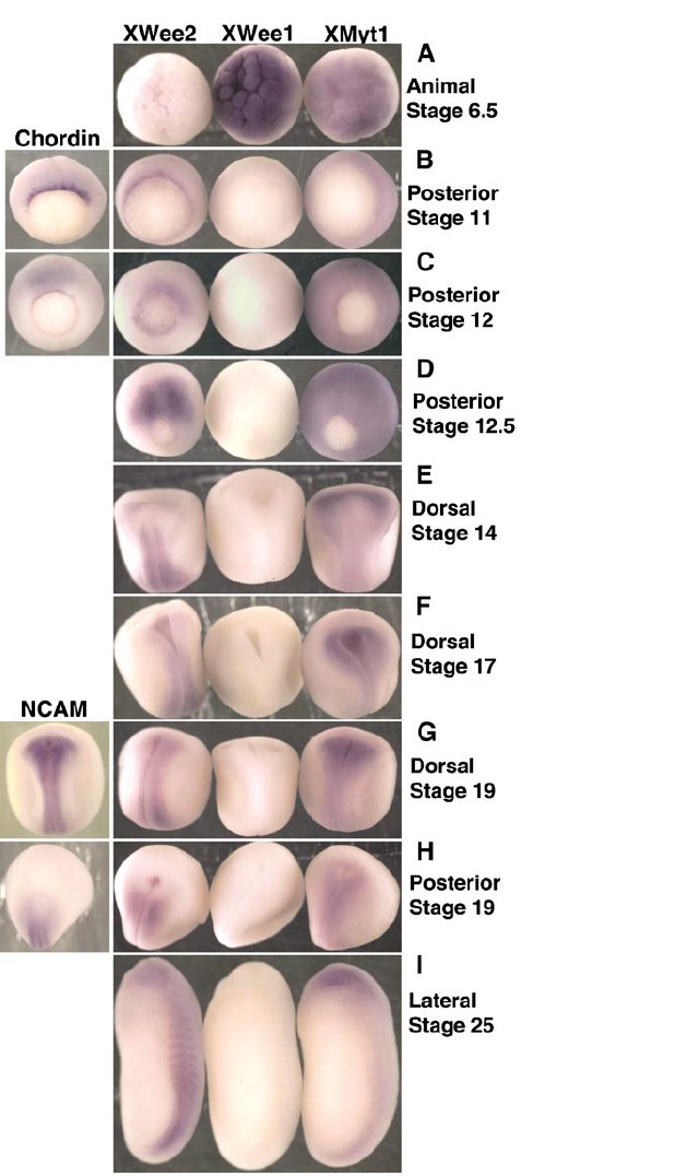
FIG. 6. Whole-mount in situ hybridization of Xenopus embryos show that each Wee kinase has a specific spatial and temporal developmental profile. Xenopus embryos were staged according to Nieuwkoop and Faber (1994), fixed, and subject to in situ hybridization for the Wee kinases (XWee2, XWee1, or XMyt1) (Aâ I), the dorsal mesoderm marker Chordin (B, C), or the neural marker NCAM (G, H). View and stages for each embryo are as indicated. See text and Materials and Methods for details.

Image published in: Leise W and Mueller PR (2002)

Copyright © 2002. Image reproduced with permission of the Publisher, Elsevier B. V.

GeneSynonymsSpeciesStage(s)Tissue
wee1wee1aXenopusThroughout NF stage 6.5animal hemisphere
wee2wee1b, xwee2XenopusThroughout gastrula stageblastopore lip
dorsal
posterior
myt1X-MyT1, xmyt1, XMyTIXenopusThroughout neurula stagedorsal
anterior neural fold
neural plate
chrdchd, chordin, chrd.1, LOC108716939, X-chordinXenopusSometime during NF stage 11 to NF stage 12dorsal
mesoderm
wee2wee1b, xwee2XenopusThroughout NF stage 13 to NF stage 21dorsal
blastopore
wee2wee1b, xwee2XenopusThroughout NF stage 25somite
dorsal
myt1X-MyT1, xmyt1, XMyTIXenopusThroughout NF stage 25eye
dorsal
anterior neural tube
myt1X-MyT1, xmyt1, XMyTIXenopusThroughout NF stage 10 to NF stage 12.5surface structure
embryo

Image source: Published

Larger Image
Printer Friendly View

Return to previous page