XB-IMG-42102
Xenbase Image ID: 42102
|
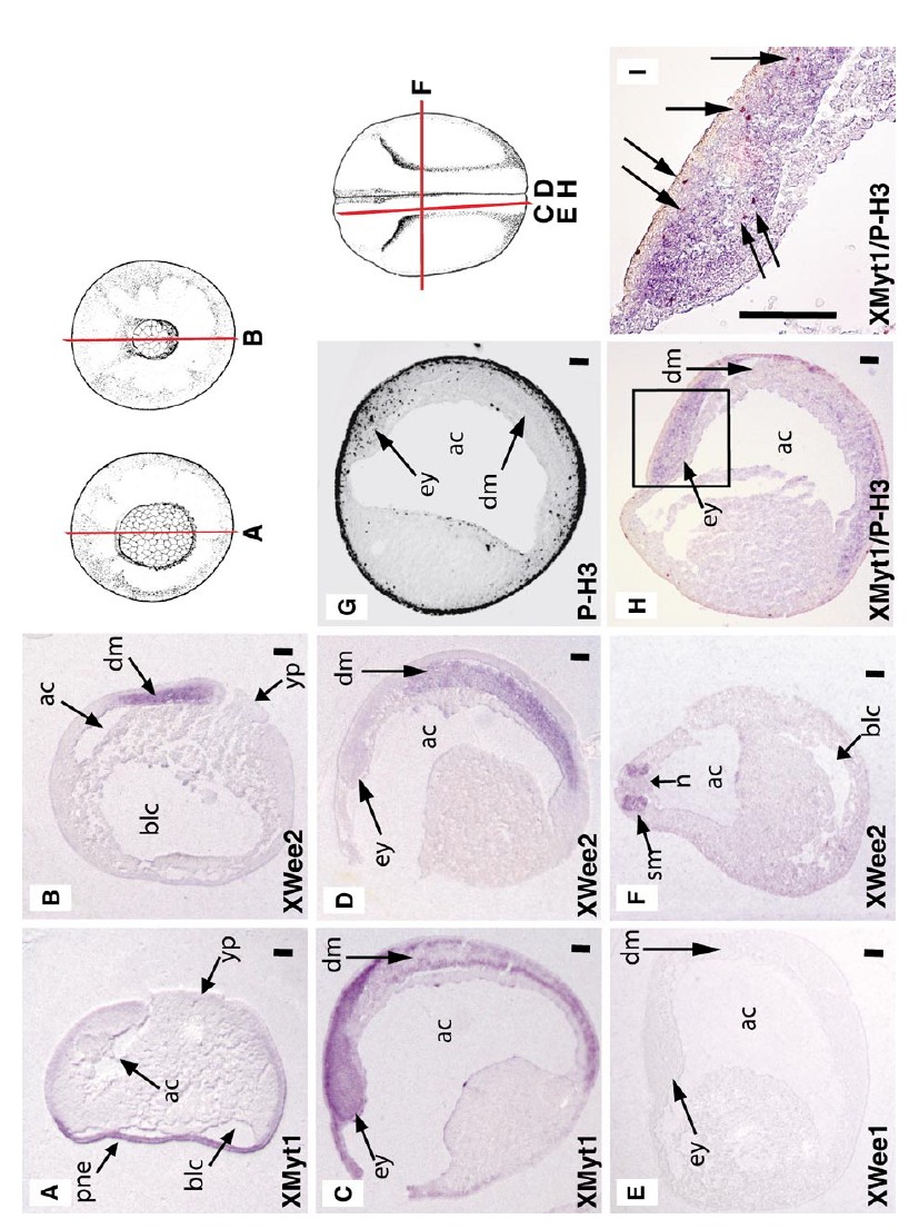
FIG. 7. XMyt1 and XWee2 are expressed in distinct germ layers of embryos undergoing gastrulation and neurulation. (A) XMyt1 is expressed in the presumptive neural ectoderm. Sagittal section of a gastrulating stage 12 embryo subjected to in situ hybridization for XMyt1. Note: the blastocoel has collapsed in this embryo. (B) XWee2 is expressed in the involuting dorsal endomesoderm. Sagittal section
of gastrulating stage 12.5 embryo subjected to in situ hybridization for XWee2. (Câ E) XMyt1 is expressed in developing neural structures, XWee2 is expressed in the dorsal mesoderm, and XWee1 is undetectable. Sagittal sections of neurulating stage 18 embryos subjected to in situ hybridization for XMyt1, XWee2, or XWee1, respectively. (F) XWee2 is expressed in the somatic mesoderm. Transverse section of neurulating stage 18 embryo subjected to in situ hybridization for XWee2. (G) Composite image of eight serial sagittal sections of a representative stage 18 embryo that was subjected to whole-mount immunocytochemistry with the PH3 antibody. Black dots indicate mitotic cells. Note the absence of mitotic cells in the involuting dorsal mesoderm. The black ring on the surface of the embryo is caused by the layering of the images and does not represent mitotic cells. (H, I) Stage 18 embryos were subjected to whole-mount XMyt1 in situ hybridization and PH3 immunocytochemistry. Saggital section of a representative embryo and enlargement of the boxed region shows that XMyt1 colocalizes to regions of high proliferation such as the eye anlage. In (I), arrows indicated mitotic nuclei (brown). For all images, positions of sections are indicated as red lines on drawings from Nieuwkoop and Farber (1994). See text for details. Scale bar, 100 m. Abbreviations: blc, blastocoel; ac, archenteron; yp, yolk plug; pne, presumptive neural ectoderm, dm, dorsal mesoderm; ey, eye anlage; sm, somite; n, notochord. Image published in: Leise W and Mueller PR (2002) Copyright © 2002. Image reproduced with permission of the Publisher, Elsevier B. V.
Image source: Published Larger Image Printer Friendly View |