XB-IMG-42890
Xenbase Image ID: 42890
|
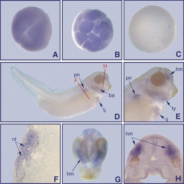
Fig. 3. Whole mount in situ hybridisation of xFoxO1. (A) Two-cell stage; (B) morula stage; (C) stage 11, vegetal view; (D) stage 35, lateral view; (E) magnification of (D); (F) transverse section showing nephric tubules; (G) anterior view on a stage 35 embryo; (H) frontal section showing head mesenchyme. Red lines in (D) indicate planes of sections shown in (F) and (H). ba, branchial arches; hm, head mesenchyme; li, liver; nt, nephric tubules; pn, pronephros; ty, thyroid gland. Image published in: Pohl BS et al. (2004) Copyright © 2004. Image reproduced with permission of the Publisher, Elsevier B. V.
Image source: Published Larger Image Printer Friendly View |