XB-IMG-43440
Xenbase Image ID: 43440
|
||||||||||||||||||||||||||||||
|
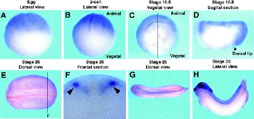
Fig. 6. WISH analysis of XDaam1 during Xenopus embryogenesis. Stages of embryos (Nieuwkoop and Faber, 1967) and views are shown above each panel. Sagittal section of embryo in C is shown in D, arrow indicates dorsal lip. Transverse sections of embryo in E are shown in F respectively, arrows indicate neural crest localization. In panels (E),(G) and (H), anterior is to the left. Image published in: Nakaya MA et al. (2004) Copyright © 2004. Image reproduced with permission of the Publisher, Elsevier B. V.
Image source: Published Larger Image Printer Friendly View |