XB-IMG-43516
Xenbase Image ID: 43516

|
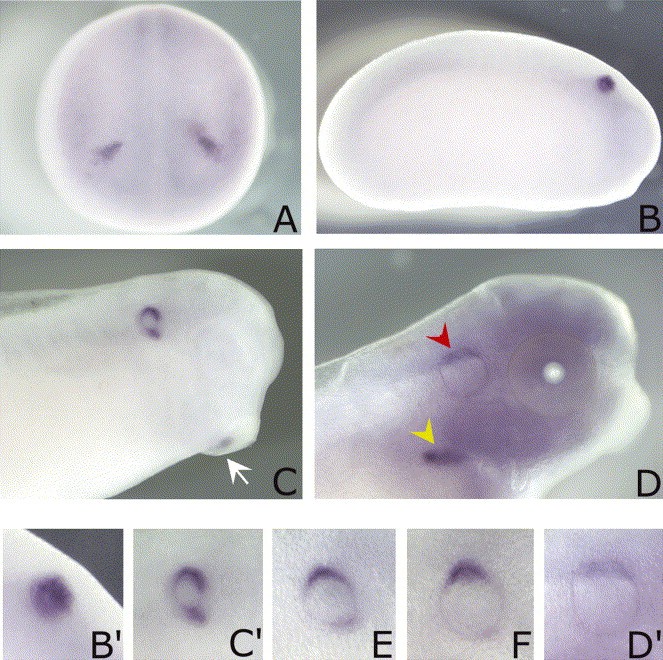
Fig. 4. Whole-mount in situ hybridization analysis of XHas3 expression pattern during Xenopus development. (A) At stage 18, XHas3 is exclusively detectable in the otic placodes. (B) A lateral view of a stage 23 embryo. (C) A stage 26 embryo. XHas3 is expressed in the otic vesicle and in the posterior half of the cement gland (arrow). (D) Stage 37/38 embryo. XHas3 is still present in the otic vesicle (red arrowhead) and appears in the pronephric glomerula (yellow arrowhead). Time course expression analysis of XHas3 in the early inner ear development. (Bâ²) Stage 23, (Câ²) stage 26: XHas3 labelling signal is higher at the dorsal and ventral sides of the otic vesicle. (E,F,Dâ²) Stage 30, 33/34 and 37/38: XHas3 mRNA gradually disappears from the ventral part of the otic vesicle walls, remaining localized at the dorsal side. Image published in: Nardini M et al. (2004) Copyright © 2004. Image reproduced with permission of the Publisher, Elsevier B. V.
Image source: Published Larger Image Printer Friendly View |
