XB-IMG-45403
Xenbase Image ID: 45403
|
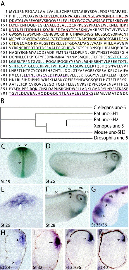
Fig. 1. (A,B) XUNC-5 protein sequence and homology. (A) Deduced XUNC-5 protein sequence. Ig domains are underlined in red, thrombospondin domains in yellow, transmembrane domain is in green, ZU5 domain is blue, DB motif is purple and the Death domain is in pink. (B) XUNC-5 homology tree. XUNC-5 shows highest homology to vertebrate UNC5H1, UNC5H2 and UNC5H3 (52, 77, 62% identity respectively). (CâK) Whole-mount and section in situ hybridization of xunc-5. (C) Dorsal view of stage 19 embryo. Anterior is up. Xunc-5 is expressed in the neural tube (arrow) and developing optic vesicles (arrowheads). (DâK) Dorsal is up and anterior is to the right. (D) At stage 26 xunc-5 transcripts are expressed around the periphery of the developing eye (asterisk) as well as in the otic vesicle (arrow), the developing brain and spinal cord. (E). Transverse section through a stage 26 embryo shows that xunc-5 transcript are confined to the dorsal spinal cord. (F) Xunc-5 expression at stage 28. Transcripts are expressed in the periphery of the developing eye (asterisk), otic vesicle (arrow), developing forebrain, midbrain, hindbrain and spinal cord. (G) Lateral view of a stage 35/36 dissected brain shows xunc-5 expression in the anterior tectum (arrow). (HâK) Transverse sections through the eye of a stage 28 (H), stage 32 (I), stages 35/36 (J) and stage 40 (K) embryo. Xunc-5 transcripts are initially expressed in both the dorsal (arrow in H) and ventral (arrowhead in H) ciliary marginal zones, and later become confined to the dorsal ciliary marginal zone (arrows in IâK). Fb, forebrain; Mb, midbrain; Hb, hindbrain; SC, spinal cord. Image published in: Anderson RB and Holt CE (2002) Copyright © 2002. Image reproduced with permission of the Publisher, Elsevier B. V.
Image source: Published Larger Image Printer Friendly View |