XB-IMG-46064
Xenbase Image ID: 46064
|
||||||||||
|
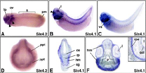
Fig. 4. Analysis of Xenopus Six4 gene expression during embryonic development. (A) Lateral view of a stage 26 embryo stained for Six4.2. The arrowheads indicate expression in the developing visceral arches. (B) Lateral view of a stage 34 embryo stained for Six4.1. The arrowhead shows the developing olfactory bulb. (C) Lateral view of a stage 39 embryo displaying widespread Six4.1 expression in the visceral arches (va). (D) Frontal view of a stage 22 embryo illustrating widespread Six4.2 in the primitive placodal thickening (ppt). (E) Transverse section of a stage 32 head cut at the level of the cement gland (cg) showing Six4.1 expression in the head mesenchyme (hm). (F) Transverse section through the head of a stage 39 embryo. Inset shows Six4.1 expression in the eye. Abbreviations: de, diencephalon; gcl, ganglion cell layer; hm, head mesenchyme; l, lens; lp, lens placode; ov, otic vesicle; pm, unsegmented paraxial mesoderm; rp, Rathke's pouch; rpe, retinal pigment epithelium; s, somites. Image published in: Ghanbari H et al. (2001) Copyright © 2001. Image reproduced with permission of the Publisher, Elsevier B. V.
Image source: Published Larger Image Printer Friendly View |