XB-IMG-46104
Xenbase Image ID: 46104
|
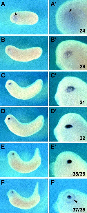
Fig. 4. Whole-mount in situ hybridization showing the expression of XL-maf. (AâF) Lateral view of whole embryos. (Aâ²âFâ²) High magnification of the eye showing XL-maf expression (AâF). (A,Aâ²) At stage 24 XL-maf is initially expressed in the PLE (arrowhead). (B,Bâ²) At stage 28 staining is detected in lens placode. (C,Câ²) Stage 31. (D,Dâ²) Stage 32. Strong signal in developing lens placode is found. (E,Eâ²) Stage 35/36. XL-maf is expressed in lens detached from ectoderm. (F,Fâ²) At stage 37/38 signal also appears in retina (arrowhead). Image published in: Ishibashi S and Yasuda K (2001) Copyright © 2001. Image reproduced with permission of the Publisher, Elsevier B. V.
Image source: Published Larger Image Printer Friendly View |