XB-IMG-47828
Xenbase Image ID: 47828
|
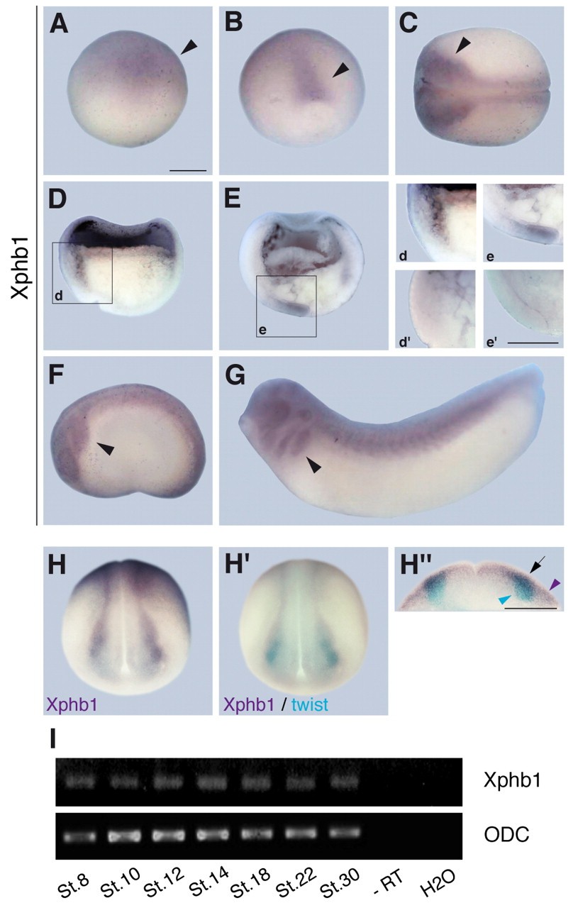
Fig. 1. Xphb1 is expressed in the presumptive cranial neural crest (CNC) region. (A-Hâ²) In situ hybridization (ISH) for Xphb1 (A-H) and Xphb1 plus twist (Hâ²,Hâ²). (A) At blastula (stage 9) Xphb1 is expressed in the dorsal animal ectoderm (arrowheads; animal pole is towards the top). (B) At late gastrula stage (stage 11.5) Xphb1 transcripts are found in the posterior dorsal area (arrowhead). (C) At neurula stage (dorsal view) Xphb1 is detected in migrating neural crest (arrowhead) and in the neural tube. (D,E) Semi-sections of stage 10 (D) and stage 11.5 (E) gastrula stage Xenopus embryos (animal pole towards the top). Insets demonstrate Xphb1 expression in the mesendoderm (d) and in the posterior neuroectoderm including mesoderm (e). No Xphb1 mRNA is detectable in comparable regions of sense control embryos (dâ²,eâ²). (F,G) From neurulation onwards (F, stage 23; G, stage 28; lateral view) Xphb1 expression is restricted to the neural crest territory (arrowheads), the eye and brain. (H-Hâ²) Double ISH at stage 18 shows that Xphb1 (H, purple) is partially co-expressed with twist (blue) in the neural crest (Hâ²). (Hâ²) Transverse section showing the colocalization of Xphb1 and twist (arrow); Xphb1 (purple arrowhead) and twist (blue arrowhead) are also expressed in non-overlapping regions. Scale bars: 400 μm. (I) RT-PCR analysis of Xphb1 expression at the indicated stages. ornithine decarboxylase (ODC) was used as an internal control. Additional controls were performed without reverse transcriptase (âRT) and without cDNA (H2O). Image published in: Schneider M et al. (2010) Copyright © 2010. Image reproduced with permission of the Publisher and the copyright holder. This is an Open Access article distributed under the terms of the Creative Commons Attribution License.
Image source: Published Larger Image Printer Friendly View |