XB-IMG-74294
Xenbase Image ID: 74294
|
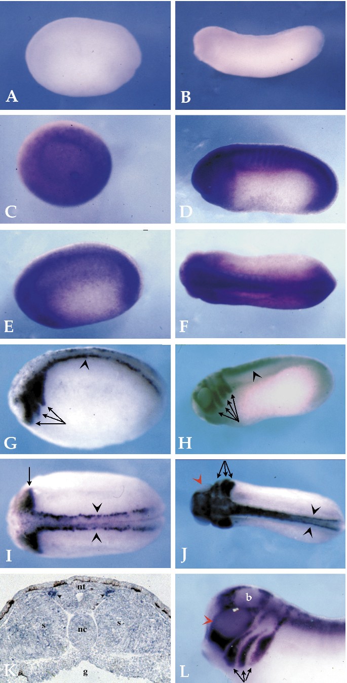
FIG. 3. Whole mount in situ mRNA hybridization of embryos at different developmental stages with xmdc9 and xmdc11a cRNA, and hybridization of a tailbud stage embryo section with xmdc11a cRNA. Albino X. laevis embryos at various developmental stages were hybridized with an xmdc9 sense probe (A, B), an xmdc9 antisense probe (C), or an xmdc11a antisense probe (G). All embryos are oriented with their anterior toward the left of the figure. A shows a lateral view of a late neurula embryo (stage 20), and B shows a tailbud embryo (stage 30). Both embryos were hybridized with the sense xmdc9 control probe. C shows that xmdc9 expression can be detected in the entire gastrula stage embryo. In a neurula embryo (E, stage 20) xmdc9 mRNA is present along the entire anteroposterior axis with a more pronounced staining on the dorsal side. At stage 25 (D), an essentially similar localization is observed in the head, the dorsal trunk structures and the tail bud. In addition, a more pronounced staining of the somites is visible. In a dorsal view of a tailbud embryo (F, stage 25), expression of xmdc9 appears strongest in dorsal structures. G and I show a lateral and dorsal view, respectively, of a late neurula embryo (stage 20) hybridized with an xmdc11a antisense probe. The most anterior signal is divided into three populations of cells (arrows) extending from the neural tube. In the trunk, a thin line of cells on either side of the neural tube express xmdc11a (black arrowheads). K shows a transverse section through the trunk of a tailbud embryo (stage 22). Two rows of xmdc11a-expressing cells (small black arrowhead) extend along the sides of the neural tube (nt). The somites (s), gut (g), and notochord structures (nc) are also indicated. In a lateral view of a tailbud stage embryo (H, stage 25), xmdc11a staining is present in four groups of cells (arrows) within the head and branchial arches. J demonstrates that expression in the same anterior structures as in H can be seen in a dorsal view of a tailbud embryo. No xmdc11a expression is detected in the optic vesicle (J, red arrowhead). In the trunk, the staining clearly appears along two stripes on either side of the midline. L depicts a magnification of the anterior region of a tailbud stage embryo. xmdc11a mRNA is localized to the four segments derived from the cranial neural crest. From posterior to anterior, these structures correspond to the posterior branchial crest, the anterior branchial crest, the hyoid crest, and the mandibular crest. The optic vesicle is marked by a red arrowhead, and the brain is indicated (b). Image published in: Cai H et al. (1998) Copyright © 1998. Image reproduced with permission of the Publisher, Elsevier B. V.
Image source: Published Larger Image Printer Friendly View |