XB-IMG-76197
Xenbase Image ID: 76197
|
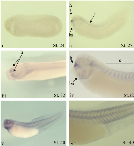
Fig. 5. Embryonic spatial expression profile of S1P genes. Whole mount in situ hybridisation with DIG-labelled RNA probe was performed on embryos from stages 6-41 for S1P genes, except for S1P4.
(B) Whole mount in situ hybridisation analysis of S1P2 expression. Lateral view of cleared em- bryos at stage 24 (i), stage 27 (ii). Dorso-lateral view of an uncleared embryo (iii) and lateral view of a cleared embryo (iv) at stage 32. Lateral view of a uncleared embryo at stage 40 (v). Detail of the stained somites of a cleared stage 40 embryo 40 (v. For the lateral views, dorsal is up and anterior is left. ba: branchial arches; h: hypophyseal anlagen; s: somites. Image published in: Massé K et al. (2010) Copyright © 2010. Image reproduced with permission of the Publisher, University of the Basque Country Press.
Image source: Published Larger Image Printer Friendly View |