XB-IMG-79699
Xenbase Image ID: 79699
|
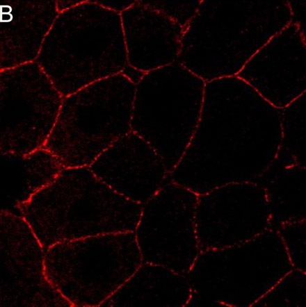
TCA-fixed animal caps were stained with an anti-C-cadherin monoclonal antibody (6B6) and visualized by Cy5-conjugated goat anti-mouse IgG. Shown here is an optical slice of a cleared animal cap imaged by LSM confocal microscope. Positive staining appears along cell-cell contacts as discrete punctae. Image published in: Tao Q et al. (2007) Copyright © 2007. Image reproduced with permission of the Publisher and the copyright holder. This is an Open Access article distributed under the terms of the Creative Commons Attribution License. Larger Image Printer Friendly View |