XB-IMG-80628
Xenbase Image ID: 80628
|
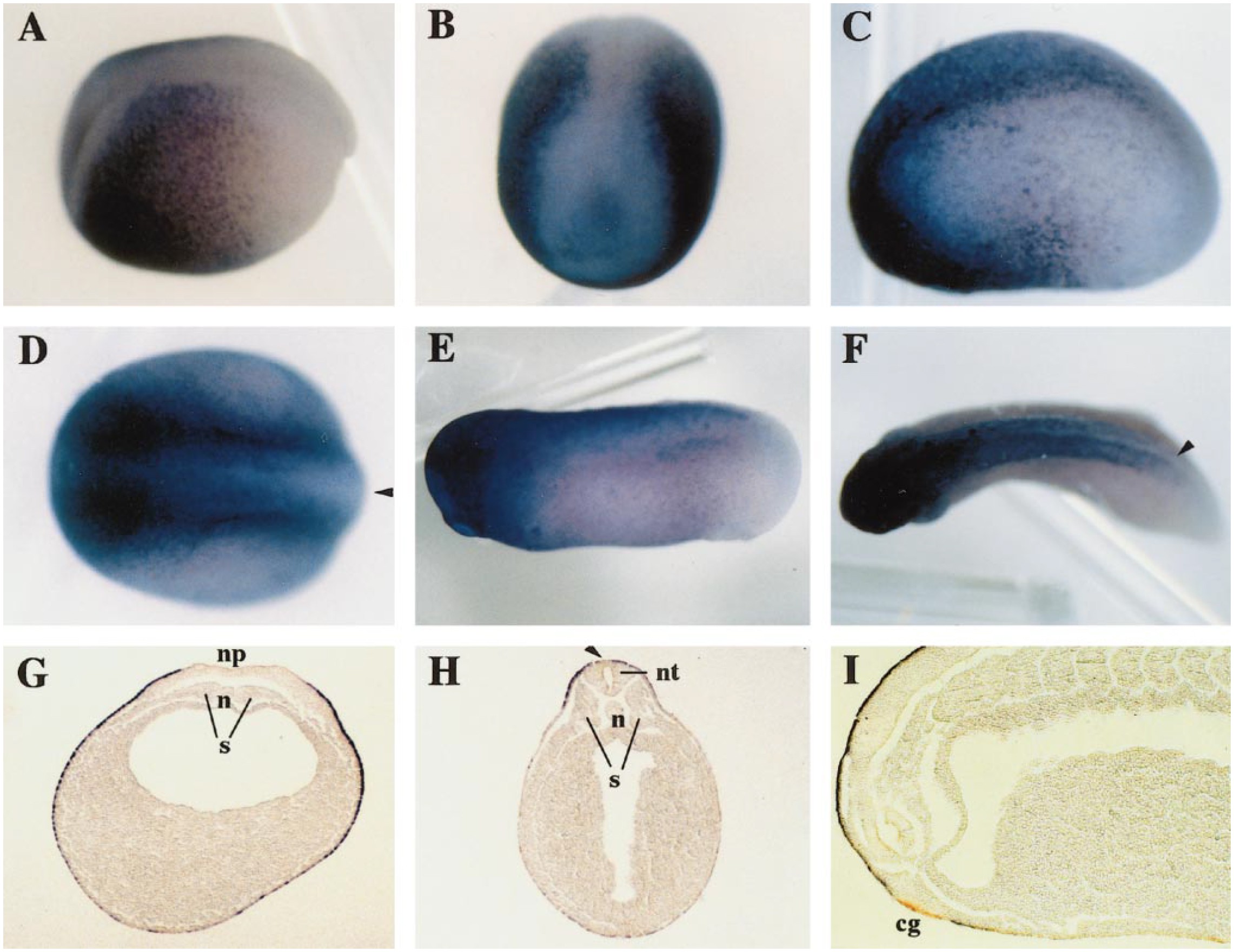
FIG. 3. Localization of Xepsin transcript in normal embryos, analyzed by whole-mount in situ hybridization. Stages are 15 (A, B), 18 (G), 20 (C, D), 22 (H, I), and 27 (E, F). (A, C, E) Lateral view. (B) Anterior view. (D, F) Dorsal view. (G) Sections of embryo after whole-mount in situ hybridization. (H) Transverse section. (I) Parasagittal section. The signal was not detected in the neural plate (np), neural tube (nt), notochord (n), somite (s), or cement gland (cg), and Xepsin expression was also absent from the midline of the most dorsal region of the epidermis (black arrowhead) (D, F, H). Image published in: Yamada K et al. (1999) Copyright © 1999. Image reproduced with permission of the Publisher, Elsevier B. V.
Image source: Published Larger Image Printer Friendly View |