XB-IMG-81354
Xenbase Image ID: 81354
|
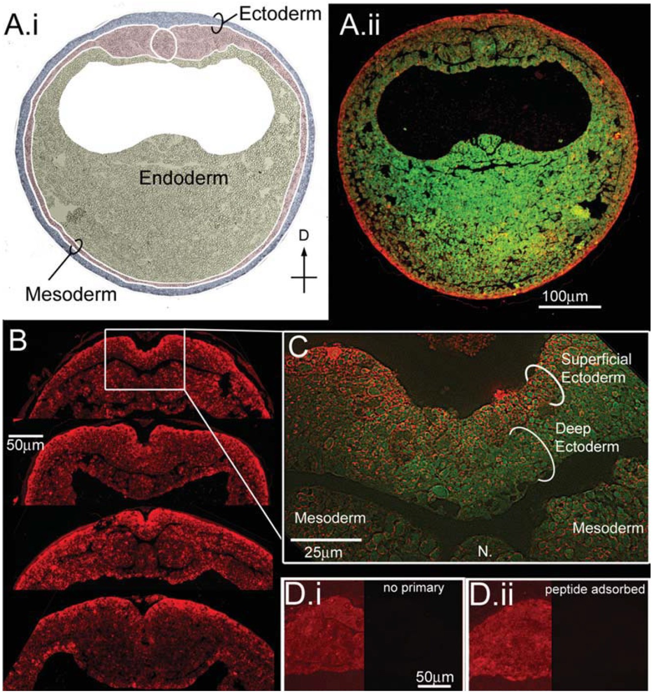
Fig. 6. Ductin is present at high levels in the
superficial ectoderm during neural plate
stages. A: Immunohistochemistry for ductin
on cross-sections from neurulating embryos.
A.i: Germ layers are indicated on the DIC
image of the section in A.ii. A.ii: Green is
autofluorescence of the section, red is ductin
signal. Ductin is ubiquitous, but stains most
strongly in the superficial ectoderm (SE) at
this stage. B: Cross-sections through the dorsal
midline showing various configurations of
the neural fold. In all sections, ductin signal is
strongest in the SE. C: Higher magnification
shows ductin concentrated in, although not
exclusively localized to, the SE. D.i: Control
section exposed to 2 Ab only. The left side of
the image has been manipulated in Photoshop
to show the position of the section; right
side of image shows the actual signal at same
exposure as B. D.ii: Control sections exposed
to peptide-adsorbed antibody; sides as in D.i. Image published in: Vandenberg LN et al. (2011) Copyright © 2011. Image reproduced with permission of the Publisher, John Wiley & Sons.
Image source: Published Larger Image Printer Friendly View |