XB-IMG-83507
Xenbase Image ID: 83507
|
||||||||||||||||||||
|
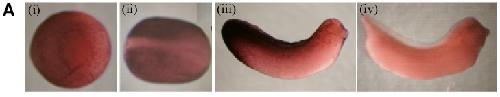
Fig. 4. Spatial expression profile of X-epilectin during development. (A) Wholemount in situ hybridisation with an X-epilectin DIG-labelled
antisense RNA probe was performed on embryos from stages 10-37. No expression was detected at stage 10 (data not shown). Expression was first detected in the epidermis from stage 12 (i). At stage 16, the staining is stronger throughout the epidermis and the neural plate is totally unstained (ii). At stage 28, X-epilectin is still expressed in the epidermis but the level of its expression is higher in the posterior part of the embryo (iii). The sense probe showed no staining pattern (iv). Image published in: Massé K et al. (2004) Copyright © 2004. Image reproduced with permission of the Publisher, University of the Basque Country Press.
Image source: Published Larger Image Printer Friendly View |