XB-IMG-87103
Xenbase Image ID: 87103
|
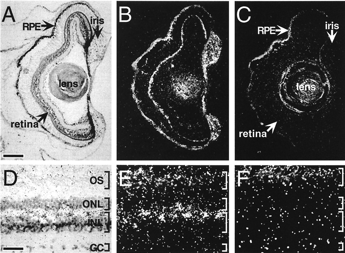
Figure 5 Bright-field and dark-field photomicrographs of melanopsin transcript distribution within ocular structures. In sections of tadpole (stages 56 and 57) (A and B) and adult (D and E) eye, melanopsin transcripts were identified in retinal cells within the outer lamina of the inner nuclear layer (INL) (A, B, D, and E). In contrast, incubation of adjacent sections with the sense RNA probe (C and F) resulted in no labeling above background. (Corresponding bright-field views are not shown.) Intense cell-specific hybridization was also observed in the iris and less intensely within the RPE. GC, ganglion cell layer; ONL, outer nuclear layer; OS, photoreceptor outer segments. [Bars = 150 μm (A) and 35 μm (D).] Image published in: Provencio I et al. (1998) Copyright © 1998. Image reproduced with permission of the Publisher.
Image source: Published Larger Image Printer Friendly View |