XB-IMG-911
Xenbase Image ID: 911
|
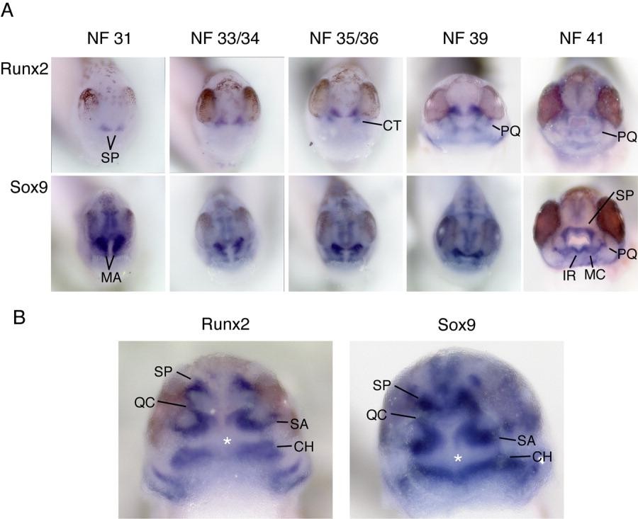
Figure 2. In situ hybridizations of runx2 and sox9 in the mandibular neural crest stream. A: Anterior views of the head showing the distribution of both transcripts over several developmental stages. Runx2 mRNA expression is first apparent in the pre-chondrogenic suprarostral plate (SP) by stage 31. Additional expression appears in the cranial trabeculae (CT) by stage 35/36 (see Fig. 3). This expression is in a subset of the mandibular arch crest (MA), which expresses sox9. The expression of sox9 mRNA is progressively restricted to co-localize with runx2. By stage 39, both transcripts overlap in the presumptive suprarostal plate (SP). The expression of runx2 decreases in the suprarostral plate by stage 41, although some expression persists in the palatoquadrate anlagen (PQ). Sox9 expression in the suprarostral plate is fused in the midline by stage 41, with continued expression in the palatoquadrate and new expression in the infrarostral (IR) and Meckel's cartilages (MC). B: Ventral views of stage 39 embryos stained with runx2 and sox9. The cement gland has been removed with a scalpel. Both transcripts are expressed in the future ceratohyal (CH) and palatoquadrate cartilages. The latter is clearly divided into a posterior sub-ocular arc (SA) and anterior quadratocranial commissure (QC), which connects to the suprarostral plate dorsally. There is a small flange of staining off of the palatoquadrate in both preparations that reveals the future Meckel's cartilage (asterisk). Image published in: Kerney R et al. (2007) Copyright © 2007. Image reproduced with permission of the Publisher, John Wiley & Sons.
Image source: Published Larger Image Printer Friendly View |