XB-IMG-149154
Xenbase Image ID: 149154
|
|
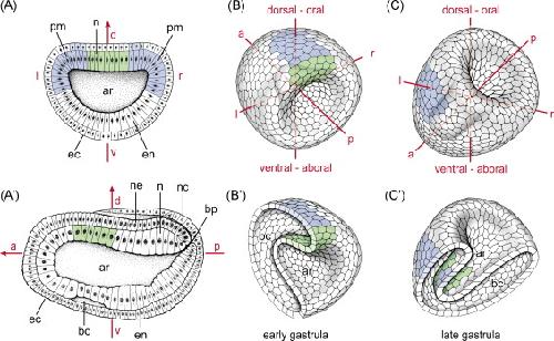
Fig. 4.
Amphioxus and sea urchin. (A) Amphioxus. Schematic drawing of a neurula stage amphioxus embryo cut transversely (A) and mid-sagittally (Aâ²). The bilateral tell-tale Nodal expression indicated in blue (A) predicts an in-between GRP in the archenteron (green in A, Aâ²). (B and C) Sea urchin. 3D schematic drawings of early (B and Bâ²) and late (C and Câ²) gastrula sea urchin embryos in whole-mount (B and C) and bisected along the dorso-ventral (Bâ²) and left-right (Câ²) axis, respectively. Proposed right half shown in (Bâ²), and oral half shown in (Câ²), Early (dorsal/aboral) symmetric Nodal organizer domain in (B) and (Bâ²) and late asymmetric expression domains in ectoderm and archenteron in (C) and (Câ²) are indicated in blue. The supposed position of a field of polarized ciliated cells is indicated in green. The conventional echinoderm body plan is inverted to account for (a) conserved left asymmetry of Nodal expression and (b) organizer gene expression on the dorsal side. Expression domains are indicated according to [9] (A), [72] (B and Bâ²) and [10] (C and Câ²). Schemes were redrawn from [9] (A) and [73] (Aâ²). a, anterior; ar, archenteron; bc, blastocoel; bp, blastoporus; d, dorsal; ec, ectoderm; en, endoderm; l, left; n, notochord; nc, neuroenteric canal; ne, neuroectoderm; pm, paraxial mesoderm; r, right; v, ventral. Image published in: Blum M et al. (2009) Copyright © 2009. Image reproduced with permission of the Publisher. Larger Image Printer Friendly View |