XB-IMG-191689
Xenbase Image ID: 191689
|
|
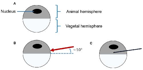
Figure 4. Process flow of the microinjection. A. Diagram of a Xenopus laevis oocyte; B. For the microinjection, the glass capillary penetrates the Xenopus laevis oocyte in the animal hemisphere, very close to the vegetal hemisphere, at an approximately 10° angle, tagged by a red arrow. C. Position of the glass capillary (shown in dark blue) during the injection. Image published in: John L and Drescher M (2018) Copyright © 2018. Image reproduced with permission of the Publisher. Larger Image Printer Friendly View |