XB-IMG-200199
Xenbase Image ID: 200199
|
|
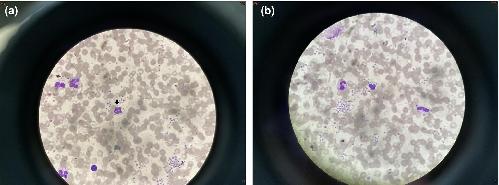
FIGURE 2
Peripheral blood smear from the proband demonstrating lymphocytosis with atypical lymphocyte morphology (Wright-Giemsa stain). (a) and (b) High magnification images (100× oil immersion) showing reactive lymphocytes with lobulated nuclei and numerous cytoplasmic vacuoles of varying sizes (indicated by arrows). Image published in: Lee CL et al. (2024) © 2024 The Author(s). Creative Commons Attribution-NonCommercial-NoDerivatives license Larger Image Printer Friendly View |