Normal Table of Xenopus development: a new graphical resource
Natalya Zahn, Christina James-Zorn, Virgilio G. Ponferrada, Dany S. Adams, Julia Grzymkowski, Daniel R. Buchholz, Nanette M. Nascone-Yoder, Marko Horb, Sally A. Moody, Peter D. Vize, Aaron M. Zorn
Development (2022) 149 (14): dev200356. https://doi.org/10.1242/dev.200356
Click here to view article at Development.
Click here to view the Zahn drawings images on Xenbase.
Click here to view the development landmarks table on Xenbase.
Click here to access the accompanying poster.
ABSTRACT
Normal tables of development are essential for studies of embryogenesis, serving as an important resource for model organisms, including the frog Xenopus laevis. Xenopus has long been used to study developmental and cell biology, and is an increasingly important model for human birth defects and disease, genomics, proteomics and toxicology. Scientists utilize Nieuwkoop and Faber's classic ‘Normal Table of Xenopus laevis (Daudin)’ and accompanying illustrations to enable experimental reproducibility and reuse the illustrations in new publications and teaching. However, it is no longer possible to obtain permission for these copyrighted illustrations. We present 133 new, high-quality illustrations of X. laevis development from fertilization to metamorphosis, with additional views that were not available in the original collection. All the images are available on Xenbase, the Xenopus knowledgebase (http://www.xenbase.org/entry/zahn.do), for download and reuse under an attributable, non-commercial creative commons license. Additionally, we have compiled a ‘Landmarks Table’ of key morphological features and marker gene expression that can be used to distinguish stages quickly and reliably (https://www.xenbase.org/entry/landmarks-table.do). This new open-access resource will facilitate Xenopus research and teaching in the decades to come.
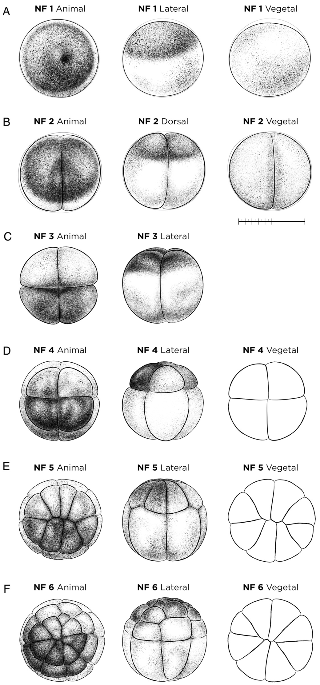
Figure 1. Cleavage-stage X. laevis embryos. (A) The fertilized egg NF stage 1. (B) NF stage 2 (two-cell stage). (C) NF stage 3 (four-cell stage). (D) NF stage 4 (eight-cell stage). (E) NF stage 5 (16-cell stage). (F) NF stage 6 (32-cell stage). Vegetal/ventral views of NF stage 4-6 (D-F) are unshaded line drawings. See Table S1 for staging landmarks. Views as indicated. Scale bar: 1 mm.
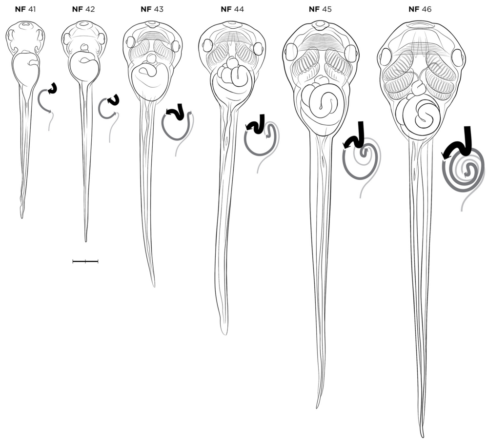
Figure 7. X. laevis embryos during gut-coiling stages, NF stages 41-46, in ventral view, alongside new gut coiling diagrams. The coiling digestive tract is depicted as three lines of varying thickness. The esophagus/stomach (thickest black line) begins anteriorly on the left side of the body at NF stage 41. As gut lengthening and coiling progresses, the stomach shifts to the right side of the body by NF stage 46. The midgut (dark-gray line) and hindgut (thin light-gray line) form a rudimentary ‘S’ shape curve by NF stage 41-42, and at NF stage 43 the midgut and hindgut have lengthened to form a ‘hairpin loop’, visible from the left side. This loop turns ventrally by NF stage 44, becoming the U-shaped apex of the future intestinal coils. Throughout NF stages 44 to 46, the midgut and hindgut continue to lengthen and loop, with the apex rotating inward to form a compact intestine with tightly wound, counterclockwise coils. Gut-coiling diagrams designed by J.G. Stages as indicated. Scale bar: 1 mm.
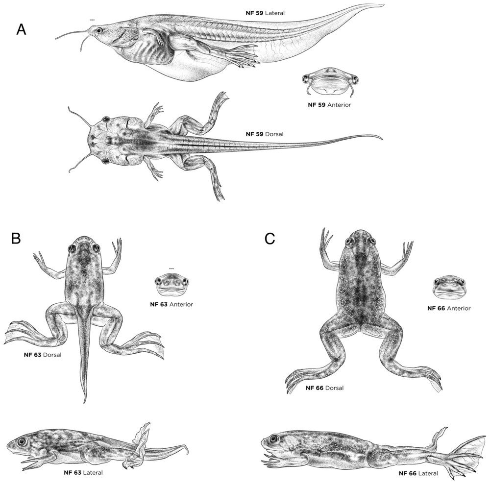
Figure 10. Prometamorphosis- and climax metamorphosis-stage X. laevis tadpoles. (A) NF stage 59. (B) NF stage 63. (C) NF stage 66. Each stage is shown in lateral, dorsal and anterior views. Ventral views are available on Xenbase. See Table S1 for more staging landmarks. Membrane removed in all embryos. Views as indicated. Scale bars: 1 mm.
Adapted with permission from The Company of Biologists on behalf of Development: Zahn et al. (2022).Normal Table of Xenopus development: a new graphical resource. Development (2022) 149 (14): dev200356. https://doi.org/10.1242/dev.200356
The Zahn Drawings are an Open Access resource distributed under the terms of the Creative Commons Attribution License CC BY-NC-4.0. You must give appropriate credit, provide a link to the license, and indicate if changes were made. You may do so in any reasonable manner, but not in any way that suggests the licensor endorses you or your use. You may not use the material for commercial purposes. To view a copy of this license, visit https://creativecommons.org/licenses/by-nc/4.0/. Contact the illustrator for commercial use.
The images or other third party material in the published article are included in the article’s Open Access Creative Commons license 4.0, unless indicated otherwise in the credit line; if the material is not included under the Creative Commons license, users will need to obtain permission from the license holder to reproduce the material. To view a copy of the articles OA license, visit http://creativecommons.org/licenses/by/4.0/
Last Updated: 2022-07-14