Focal Impact Model of Traumatic Brain Injury in Xenopus
A Focal Impact Model of Traumatic Brain Injury in Xenopus Tadpoles Reveals Behavioral Alterations, Neuroinflammation, and an Astroglial Response
Sydnee L Spruiell Eldridge, Jonathan F K Teetsel, Ray A Torres, Christina H Ulrich, Vrutant V Shah, Devanshi Singh, Melissa J Zamora, Steven Zamora, Amy K Sater
Int J Mol Sci. 2022 Jul 8;23(14):7578. doi: 10.3390/ijms23147578.
Click here to view article at International Journal of Molecular Sciences.
Click here to view article on Pubmed.
Click here to view article on Xenbase.

Abstract
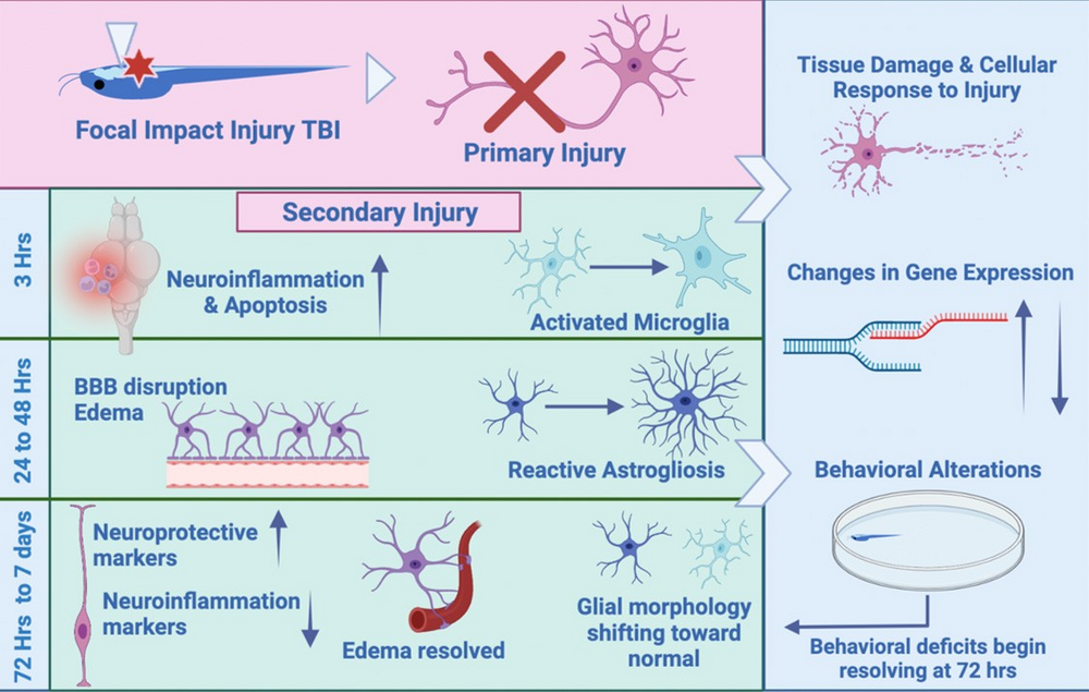
Traumatic Brain Injury (TBI) is a global driver of disability, and we currently lack effective therapies to promote neural repair and recovery. TBI is characterized by an initial insult, followed by a secondary injury cascade, including inflammation, excitotoxicity, and glial cellular response. This cascade incorporates molecular mechanisms that represent potential targets of therapeutic intervention. In this study, we investigate the response to focal impact injury to the optic tectum of Xenopus laevis tadpoles. This injury disrupts the blood-brain barrier, causing edema, and produces deficits in visually-driven behaviors which are resolved within one week. Within 3 h, injured brains show a dramatic transcriptional activation of inflammatory cytokines, upregulation of genes associated with inflammation, and recruitment of microglia to the injury site and surrounding tissue. Shortly afterward, astrocytes undergo morphological alterations and accumulate near the injury site, and these changes persist for at least 48 h following injury. Genes associated with astrocyte reactivity and neuroprotective functions also show elevated levels of expression following injury. Since our results demonstrate that the response to focal impact injury in Xenopus resembles the cellular alterations observed in rodents and other mammalian models, the Xenopus tadpole offers a new, scalable vertebrate model for TBI.

Figure 1
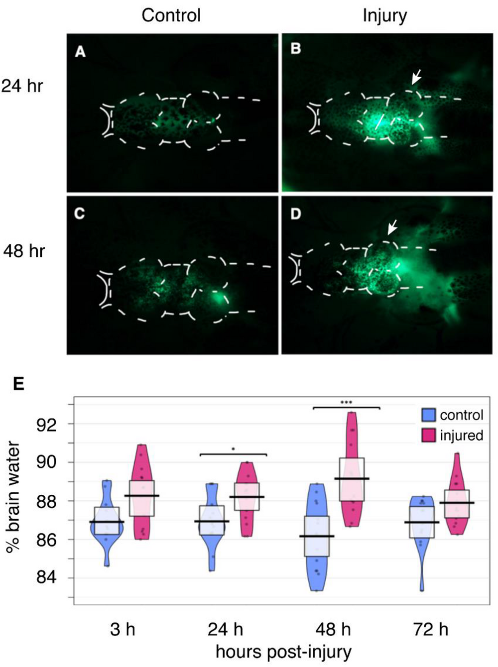
Focal Impact Injury leads to Edema and Disruption of BBB integrity. Sham tadpoles were anesthetized and transferred temporarily to the injury cradle prior to intraventricular injection with sodium fluorescein (NaF) either 24 h post-injury (h.p.i.) (A) or 48 h.p.i (C). Injured animals were anesthetized and then subjected to focal impact injury at the optic tectum (white arrows). Following either 24 h (B) or 48 h (D) of recovery post-injury, tadpoles were given an intraventricular injection with 10 nL of 1 μg/mL NaF; diffusion of the tracer was visualized using fluorescence microscopy. Arrows in (B,D) show the site of injury. (A–C) White dotted lines indicate the outer borders of the brain. Images are representative of n = 6 animals per treatment group for each time point. (E) * p < 0.05; *** p < 0.001.

Figure 6
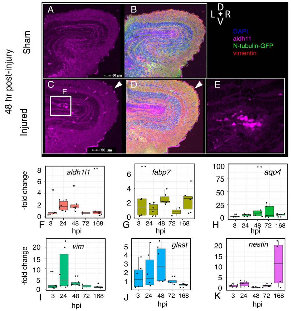
Persistence of altered astrocyte morphology and expression of astroglial genes following injury. Visualization of astrocytes (aldh1l1-positive cells, magenta), neurons (N-tubulin-GFP), radial glia (vimentin-positive, red) in sham (A,B) and injured (C–E) 48 h after injury. Amoeboid astrocytes accumulate near the ventricular layer (C,E). Quantitative RT-PCR assays for expression of astroglia-associated genes Aldehyde dehydrogenase 1 family member 1 (aldh1l1) (F), fatty acid-binding protein 7 (fabp7) (G), aquaporin4 (aqp4) (H), vimentin (vim) (I), excitatory amino acid transporter 1 (glast) (J), and nestin (nes) (K). RNA was isolated from sham and injured midbrains at the intervals shown; injured values are normalized to sham-treated controls. Plots show individual data points and a box showing the mean and 95% confidence intervals. n ≥ 6 experiments.

Figure 10
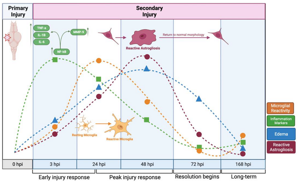
A timeline of response to focal impact injury. The response to focal impact injury begins with the release of inflammatory cytokines, followed by microglial activity, astrocyte reactivity, and edema. Within 48 h, each of these activities has peaked, and all have subsided to near-baseline levels after one week (This image was made using BioRender (Suite 200, Toronto, ON, M5V 2J1)).
Adapted with permission from MDPI on behalf of The International Journal of Molecular Sciences: Spruiell Eldridge et al. (2022). A Focal Impact Model of Traumatic Brain Injury in Xenopus Tadpoles Reveals Behavioral Alterations, Neuroinflammation, and an Astroglial Response. Int J Mol Sci. 2022 Jul 8;23(14):7578. doi: 10.3390/ijms23147578.
This work is licensed under a Creative Commons Attribution 4.0 International License. The images or other third party material in this article are included in the article’s Creative Commons license, unless indicated otherwise in the credit line; if the material is not included under the Creative Commons license, users will need to obtain permission from the license holder to reproduce the material. To view a copy of this license, visit http://creativecommons.org/licenses/by/4.0/
Last Updated: 2022-08-24