XB-IMG-130142
Xenbase Image ID: 130142
|
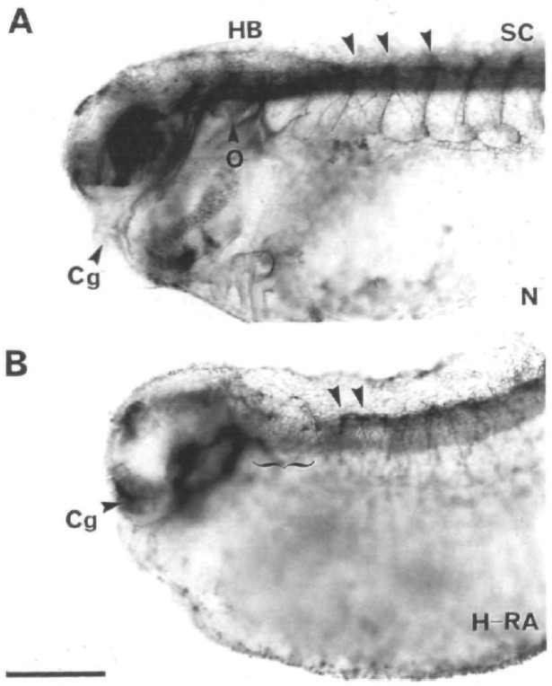
Fig. 7. Expression of HNK-1 by Rohon-Beard neurons in
normal and RA-treated embryos. Panel A shows the
pattern of expression of the HNK-1 antigen in wholemount
preparations of tadpole stage (stage â38-40)
embryos. mAb HNK-1 labels a variety of structures in the
CNS including the cell bodies of Rohon-Beard neurons in
the dorsal spinal cord (arrowheads). Panel B shows the
labelling pattern of HNK-1 in the spinal cord of an embryo
treated with high concentrations of RA. The inhibition of
normal anterior development is not accompanied by an
anterior extension of the domain over which Rohon-Beard
neurons appear (bracket). Cg, cement gland; HB,
hindbrain; o, otic vesicle; RA, embryo treated with high
(H-RA) concentration of RA; SC, spinal cord. Scale
bar=0.5mm. Image published in: Ruiz i Altaba A and Jessell TM (1991) Copyright © 1991. Image reproduced with permission of the Publisher and the copyright holder. This is an Open Access article distributed under the terms of the Creative Commons Attribution License.
Image source: Published Larger Image Printer Friendly View |