XB-IMG-129902
Xenbase Image ID: 129902
|
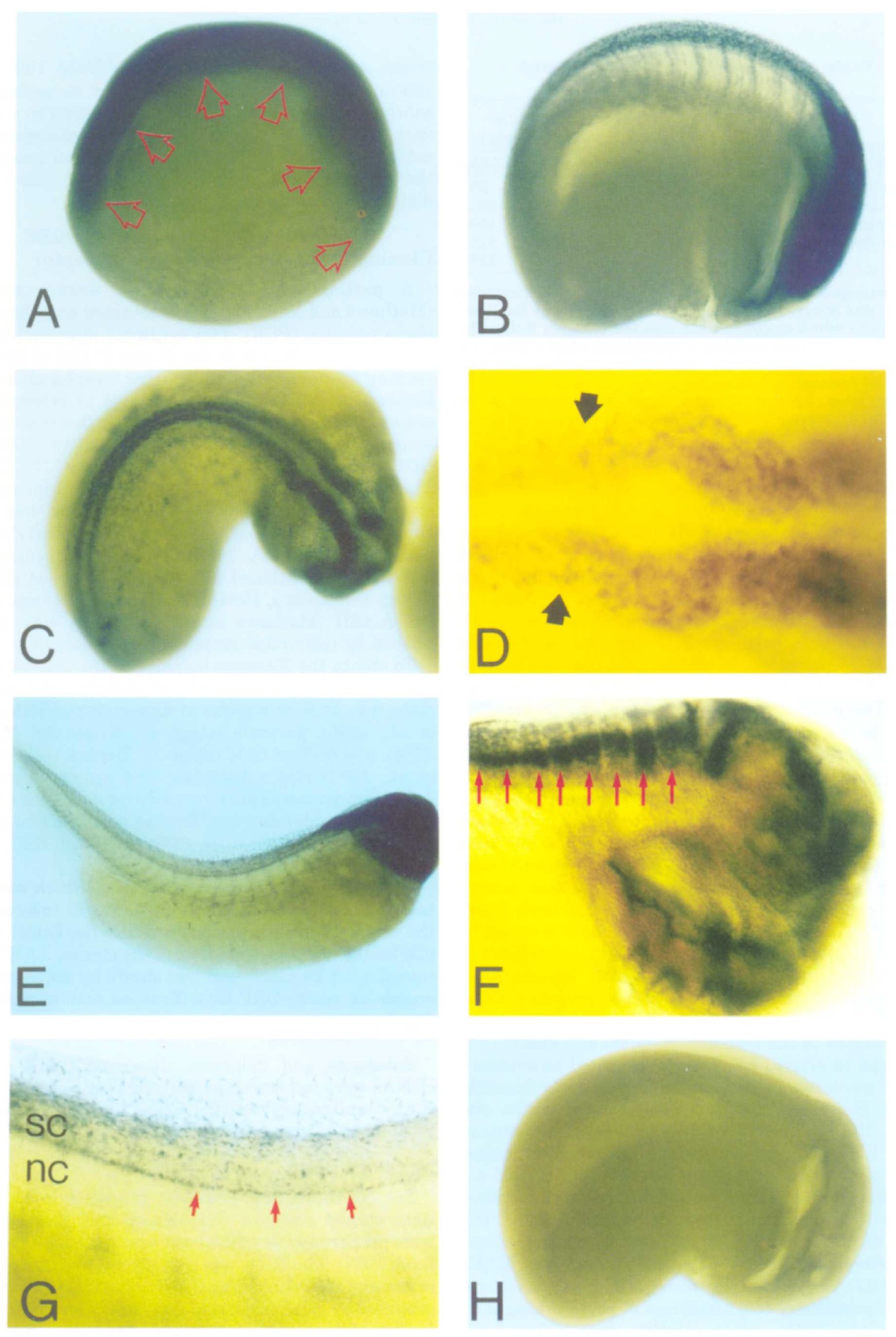
Fig. 4. Spatial distribution of the XAR1 transcript during neurogenesis
detected by whole mount in-situ hybridization. A: At neurula stage 15 the
XARl transcript is detected in neuroectoderm. Arrows delineate the neural
plate. 6: Stage 21 neurula. XARl RNA is detected in the brain, eyes,
and spinal cord. C: Dorsal view of a stage 23 embryo. Strong expression
is detected in the CNS on each side of midline. D: Close-up view of XARl
RNA in the anterior CNS. The arrows point to the hindbrain-spinal cord
junction. E: Stage 35 tadpole. Most of the transcript is localized in the
head of the embryo. F: Close-up view of the head of the same embryo.
Arrows point to the rhornbomeres of the hindbrain that show strong expression.
Expression can also be seen at the midbrain-hindbrain and
midbrain-forebrain junction, anterior wall of the forebrain, the eye (outside
of this focal plan), and mandibular arches and pharyngeal pouches. G:
Expression of XARl in the spinal cord of the same embryo as shown (E).
The RNA is expressed in cells aligned in the ventral side of the spinal
cord (arrows). Some cells in the midtrunk region of the dorsal fin are also
stained. H: A stage 24 neurula stained with XARl sense probe as a
control. All embryos presented are albino. In all cases, the dorsal side is
up and anterior is to the right. Panel (F) and (G) were taken under differential
interference contrast optics, all other panels were bright light
optics. sc = spinal cord, nc = notochord. Image published in: Hemmati-Brivanlou A et al. (1992) Copyright © 1992. Image reproduced with permission of the Publisher, John Wiley & Sons.
Image source: Published Larger Image Printer Friendly View |