XB-IMG-133816
Xenbase Image ID: 133816
|
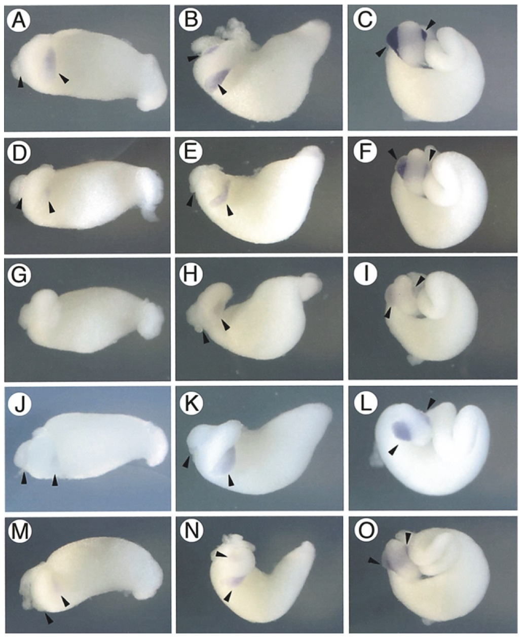
Fig. 5. Expression pattern of
pancreatic genes. Embryos at
three stages were examined by in
situ hybridization using specific
probes for each clone. CEL was
expressed in dorsal and ventral
pancreas rudiments (arrowhead)
at stage 40 and continued after
stage 42 (A,B,C: stages 40, 42
and 44, respectively). PE2 (D,E,F:
stages 40, 42 and 44, respectively),
PDIp (J,K,L: stages 40, 42
and 44, respectively) and DNaseI
(M,N,O: stages 40, 42 and 44,
respectively) were detected as
weak signals at stage 40 and
strong signals at stage 42. PP11
(G,H,I: stages 40, 42 and 44,
respectively) was barely detectable
at stage 40 but expression
increased by stage 42. Image published in: Sogame A et al. (2003) Copyright © 2003. Image reproduced with permission of the Publisher, John Wiley & Sons.
Image source: Published Larger Image Printer Friendly View |